Biotech Strategy Blog

Commentary on Science, Innovation & New Products with a focus on Oncology, Hematology & Cancer Immunotherapy

Posts from the ‘Alzheimer’s’ category

It’s a fact of human life that we lose physical and mental function as we get older. In the information age that we currently live in, this translates into a decline in our ability to function and perform the activities of daily living. Can we halt or delay age-related memory loss?

Min Wang and colleagues from Yale University School of Medicine in the August 11 issue of Nature, have published some elegant research that suggests we may be able to, at some point in the future.

It’s important to distinguish the cognitive loss associated with normal ageing from that associated with dementias such as Alzheimer’s disease where major changes to the brain structure and function occur. The Yale researchers accomplished this by using aged monkeys that have a highly developed prefrontal cortex (PFC), the part of the brain associated with working memory. Monkeys, unlike humans, do not develop age-related dementias!

Working memory that allows you to keep things “in mind” e.g. where you put the car keys down, relies on a network of pyramidal neurons in the dorsolateral PFC that excite each other.

The strength of this excitatory network depends on the neurochemical environment e.g. elevated cAMP signaling reduces nerve firing. Wang and colleagues reversed the age-related decline in PFC activity by restoring an optimal neurochemical environment. Through a series of experiments they found that:

The memory-related firing of aged DELAY neurons was partially restored to more youthful levels by inhibiting cAMP signalling, or by blocking HCN or KCNQ channels.

These findings reveal the cellular basis of age-related cognitive decline in dorsolateral PFC, and demonstrate that physiological integrity can be rescued by addressing the molecular needs of PFC circuits.

This research, although preliminary and based on animal models, is promising. It offers the hope that in the future we may be able to reverse or slow-down the age-related memory loss and cognitive defects we would otherwise experience.

Many biotechnology and pharmaceutical companies are focusing on Alzheimer’s disease as a target. What this research suggests is that developing therapies that may delay or slow-down age-related memory decline could also be a valid target for drug development, with a significant market opportunity.

ResearchBlogging.orgWang, M., Gamo, N., Yang, Y., Jin, L., Wang, X., Laubach, M., Mazer, J., Lee, D., & Arnsten, A. (2011). Neuronal basis of age-related working memory decline Nature, 476 (7359), 210-213 DOI: 10.1038/nature10243

BIO-2011-Interational-Convention-Washington-DC

I am excited to be attending, for the first time, the Biotechnology Industry Organization (BIO) international convention that takes place in Washington DC in just over a week’s time from Monday June 27 to Thursday, June 30th.

This meeting has something for everyone interested in the biotechnology industry whether it be deal making, partnering, licensing, drug discovery or personalized medicine. There are 16 specialized tracks where industry experts provide insight and best practices.

In addition, there are numerous networking and social events plus an exhibit hall that showcases the world’s biotech regions and how they are promoting innovation.

At meetings where there are parallel sessions, I apply “the law of two feet” (thanks to Podcamp for this) that says if you are not getting what you want from the session, it’s OK to walk out and go to another one.

My top 10 sessions at BIO reflect my personal interests in innovation, science and new product development:

Tuesday June 28

  • How will we afford Personalized Medicines?
  • The Biomarkers Consortium: Facilitating the Development and Qualification of Biological Markers
  • Personalized Oncology: The emergence of Personalized Medicine Strategies in Oncology Clinical Development and Deal Making
  • Navigating the New Law on Licensing Biosimilars

Wednesday June 29

  • Lessons from a Mature Public-Private Partnership. The Alzheimer’s Disease Neuroimaging Initiative
  • Emerging Markets. The Future of Growth for Biologics?
  • The Role of Imaging Biomarkers in Early Phase CNS Drug Development
  • The Promise of MicroRNA-based Therapeutics in Cancer

Thursday Jun 30

  • After the Fall. Venture Capital and the Biotech Funding Landscape
  • Regulatory Issues for Tissue Engineered Products

If you have plans to be at BIO 2011 do say hello after one of the sessions or receptions. You can reach me at the meeting via twitter (@3NT).  See you in DC!

Follow 3NT on Twitter

1 Comment

Due to the pressure of other commitments, I only had the pleasure of attending the annual meeting of the Association for Research in Vision and Ophthalmology (ARVO) for two days, but one of my key take home messages from the meeting is how we can use the eye as a window into the brain.  This is particularly relevant to Alzheimer’s research.

ARVO researchers at a lunchtime workshop that I attended asked the question of what can we learn from shared disease mechanisms in age-related macular degeneration (AMD), Alzheimer’s Disease (AD) and Glaucoma to devise therapies of the future?

What I learnt in the introduction by Nicholas Bazan from LSU Health Sciences is that both AD and AMD are both multifactorial, genetically complex, progressive, late-onset neurodegenerative conditions.  Common features include:

  1. Age-related neurodegeneration
  2. Amyloid precursor protein mis-processing
  3. Non-resolving inflammatory response
  4. Selective apoptotic cell death

Researchers in the workshop presented early experimental findings.

Catherine Bowes Rickman from Duke presented data that showed anti-amyloid immunotherapy blocks retinal pigment epithelium (RPE) damage and visual function defects in an AMD-like mouse model.  Interesting questions were raised as to whether mouse Aß aggregates differently to human, so is this a good model?

Adriana Di Polo from the University of Montreal discussed Glaucoma and AD: common neurodegenerative pathways and therapeutic targets. It was interesting to note that high rates of visual abnormalities, including glaucoma, have been reported in AD patients, but causality has not been established. Neuronal loss in both glaucoma and alzheimer’s disease occurs via common cell death processes including altered metabolism of Amyloid Precursor Protein (APP) and Aß.

What Di Polo highlighted in her talk was the potential to use therapies effective in one disease to treat the other e.g. galantamine is approved for treatment of mild to moderate AD symptoms.  Because it crosses the retinal-brain barrier and has high bioavailability, she presented results using this in an animal model of glaucoma.

Her conclusion was that “therapeutic modalities that promote neuroprotection in AD may be useful in glaucoma and vice versa.”

The third speaker of this fascinating workshop was Ian Trounce from Melbourne, who challenged the Amyloid theory of AD. His hypothesis was that sAPPα may trigger oxidative stress in mitochondria and be the problem. He discussed the increasing acceptance/overlap in pathologies between Parkinson’s and AD.  He presented data that sAPPα overexpression protects retinal ganglion cells (RGC) from rotenone via PI3K-AKT activation.

Critical feedback on the three presentations was provided by Guy Eakin of the American Health Assistance Foundation (AHAF) and Imre Lengyel from UCL.

As Dr Lengyel succinctly notes in his UCL Institute of Ophthalmology bio:

“It appears that the development of age related macular degeneration (AMD) and Alzheimer’s disease (AD) share similar histopathology, vascular risk factors and genetic predisposition. In addition, the development of AMD appears to use similar or identical steps on the cellular and molecular levels to AD: vascular damage, oxidative stress, inflammation, extracellular protein and peptide degradation or deposition, and the role for lipids and trace elements (especially zinc) in the degenerative process are amongst the many common features. Furthermore, amyloid beta peptides are an integral part of drusen (the hallmark lesion in AMD) and their formation might be similar to plaque formation in AD.”

I applaud ARVO for looking at how the eye can be used as a window into the brain. It raises the intriguing prospect that research on AMD may not only help understand the cause of AD, but that the eye may serve as an experimental model for future new treatments. Collaboration between Opthalmology and Alzheimer’s researchers is something I expect and hope we will see more of.


The highlight of the recent Association of Health Care Journalists (AHCJ) annual meeting in Philadelphia (Health Journalism 2011) for me was the presentation by Kacy Cullen from the Center for Brain Injury and Repair in the Department of Neurosurgery at the University of Pennsylvania.

© Kacy Cullen, University of Pennsylvania

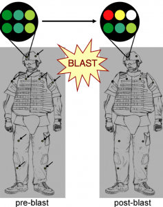
Dr Cullen presented his research on blast-induced traumatic brain injury (bTBI) and the development of a nanomaterial containing photonic crystals that change color upon exposure to blast pressure.

In the same way that a radiation dosimeter badge records exposure to cumulative radiation for a hospital worker, so a helmet-mounted color badge would change color based on a soldier’s exposure to blast pressure; a common occurrence with improvised explosive devices (IED).

In a paper published in NeuroImage, Cullen and colleagues describe in detail a blast-injury dosimeter (BID) made from photosensitive polymers that is like a colored sticker.  This nanomaterial contains microscopic, diamond-like photonic crystals, whose ability to refract light is damaged in a precise way by the pressure from explosive blasts.

The result is a change in color that is related to the degree of pressure and blast intensity. What’s more because the photonic crystals are structurally damaged by the blast, further exposure leads to more widespread microstructural alterations and a further change in color.  In essence, the crystals have a memory for cumulative blast exposure.

Why is this important?

Many soldiers are exposed to blasts, but show no overt symptoms of traumatic brain injury.  Research has shown that repeated hits to the helmet of a football player can lead to brain injury without the obvious signs of a concussion.  Traumatic brain injury as a result of repeated exposure to blasts may also lead to mild cognitive impairment and the possibility of increased risk for dementia, Alzheimer’s disease later in life.  This has been seen in NFL players.

The research by Cullen and colleagues is still in the early stages of development.  In their paper they acknowledge some of the next steps such as calibrating the color changes to levels of blast exposure, and correlating these with traumatic brain injury.  Any blast injury dosimeter will also need to be field tested.

However, this work is promising and an example of how nanotechnology may impact the detection and diagnosis of those soldiers at risk of traumatic brain injury.

War related scientific research often leads to civilian applications. In the future, I could see nanotechnology stickers that change color with cumulative impact on the helmets of NFL, college or high school football players.

You can read more about this innovative research on how color changing photonic crystals detect blast exposure in the journal NeuroImage.

Update June 30, 2011

If you are interested in the exciting and innovative research being undertaken by Kacy Cullen and his team, there is now a website for The Cullen Laboratory and their work on Neural Engineering in Neurotrauma.

ResearchBlogging.orgCullen, D., Xu, Y., Reneer, D., Browne, K., Geddes, J., Yang, S., & Smith, D. (2011). Color changing photonic crystals detect blast exposure NeuroImage, 54 DOI: 10.1016/j.neuroimage.2010.10.076

Changes in brain structure, function and molecular processes occur several years before clinical symptoms of Alzheimer’s disease (AD) become apparent.

The big question then, is can you detect patients who are cognitively normal, but will go on to develop AD before they show symptoms, i.e. pre-symptomatic patients?  The answer is “Yes” according to results published in the April 19, 2011 issue of Neurology by Brad Dickerson and colleagues.

In this small study, the team of researchers from two centers (Massachusetts General Hospital and Rush University in Chicago) followed a small sample of cognitively normal (CN) subjects over time with magnetic resonance imaging (MRI) and then sought to identify what structural changes had taken place in those subjects who were initially cognitively normal, but went on to develop AD, on average 11.1 years later.

The researchers found that changes in brain cortical thickness were associated with AD:

AD-signature cortical thinning in CN-AD converters in both samples was remarkably similar, about 0.2 mm (p < 0.05)

They concluded that:

By focusing on cortical regions known to be affected in AD dementia, subtle but reliable atrophy is identifiable in asymptomatic individuals nearly a decade before dementia, making this measure a potentially important imaging biomarker of early neurodegeneration.

Some of the limitations of this research and questions that come to mind are:

  • Small sample size: only 8 individuals who developed AD and 25 in the cognitively normal control group.
  • Reproducibility: the 0.2mm difference seen is small and the extent to which other centers may be able to reproduce this measurement is uncertain
  • Accuracy of detection: in any screening tool the issue of false positives and negatives arises i.e. in a larger sample size will there be a margin for error that results in some people being included in the pre-symptomatic AD group, when they may be normal?  Also will the proposed measurement remain valid in a large population of patients with other disease symptoms and chronic illnesses?
  • Validity of biomarker: are the changes in cortical thickness causally linked to AD or just an incidental correlation i.e. is this a valid biomarker?

Brad Dickerson in the excellent Neurology podcast available with this publication clearly sees this currently as a research tool, especially given the requirement for considerable computer power to make these types of cortical measurements in the brain.  The podcast interview is well worth listening to.

The MRI biomarker proposed by Dickerson is therefore not something that is really applicable to screen the general population at the moment.

However, the promise from this and other biomarker research is that at some point in the not too distant future we will be able to detect those at risk of developing AD. Those patients could then be given neuroprotective drugs that may delay the onset of the clinical symptoms of AD such as memory loss and cognitive impairment.

Biomarkers that identify those at risk of developing AD will also be useful as inclusion and screening tools for clinical trials of drugs aimed at slowing disease progression in pre-symptomatic patients.

Alzheimer’s disease has been called “The challenge of the Second Century,” we still have a long way to go before this is overcome.

Story Source:  BBC Health

ResearchBlogging.orgDickerson, B., Stoub, T., Shah, R., Sperling, R., Killiany, R., Albert, M., Hyman, B., Blacker, D., & deToledo-Morrell, L. (2011). Alzheimer-signature MRI biomarker predicts AD dementia in cognitively normal adults Neurology, 76 (16), 1395-1402 DOI: 10.1212/WNL.0b013e3182166e96

Earlier this month the Michael J Fox Foundation (MJFF) announced that Vancouver based Allon Therapeutics had been able to improve motor function and brain pathology in a mouse model of Parkinson’s disease (PD).

MJFF funded this research with Allon Therapeutics. The preclinical study results are published in the Journal of Molecular and Cellular Neuroscience.

What makes this data interesting is that it adds further support to the potential efficacy of the company’s lead product, davunetide, in a wide range of neurodegenerative disorders.

Davunetide (AL-108) is a microtubule-interacting peptide based on an eight amino acid sequence, Asn-Ala-Pro-Val-Ser-Ile-Pro-Gln, single letter code NAPVSIPQ (NAP) derived from activity-dependent neuroprotective protein (ADNP). It has been shown to have neuroprotective properties.

Davunetide can be administered by IV or intranasally and crosses the blood/brain barrier. It is effective at promoting neurite growth, restoring transmission between nerve cells and untangling some of the damage seen in neurodegenerative disorders such as Alzheimer’s disease. References to the scientific publications and mechanism of action can be found on the Allon Therapeutics website.

Currently it is being developed for Alzheimer’s disease (AD), schizophrenia cognitive impairment and frontotemporal dementia (FTD). Davunetide is in a phase 3 clinical trial for progressive supranuclear palsy (PSP), a subtype of FTD.

The company’s strategy is to pursue a fast-track to market in a small indication such as PSP. This is makes a lot of sense for a small biotechnology company with limited funding.  Successful approval in PSP will significantly increase the value of the company and improve the terms of any future licensing/partnering deals.

The hope for davunetide is that it will prevent disease progression in disorders such as AD and provide neuroprotective prophylaxisis prior to surgery that carries a high risk of memory loss e.g. heart bypass and coronary artery graft surgery (CABG).

While davunetide may not be a cure for AD, being able to slow down disease progression is something that has considerable value.  Given that new imaging biomarkers are likely to provide the opportunity to detect AD much earlier, the market opportunity for early treatment is set to increase.

Many families and caregivers would welcome a drug that delays further cognitive decline and memory loss in their loved ones.

On the basis of the promising preclinical results, I think we can expect to see further clinical research on davenutide in Parkinson’s Disease.

My theme for blog posts this week has been the diagnosis and detection of Alzheimer’s Disease, a therapeutic area I was first introduced to while working as a Global Project Director at the Canadian CRO, CroMedica before it was acquired by PRA. The then CEO of CroMedica, Erich Mohr Ph.D is now Chairman and CEO of MedGenesis Therapeutix Inc. in Victoria, BC.

This privately held biopharmaceutical company is working on developing new products for neurological diseases and the treatment of Parkinson’s Disease, Glioblastoma Multiforme (GBM) and Intractable Epilepsy. I have added MedGenesis to my list of emerging biotechnology companies to watch, and look forward to writing further as their pipeline develops.

Which brings me back to Alzheimer’s disease (AD), an area which I think will touch many of us as we and our parents become older. Last week, I was visiting my elderly mother in England who struggles to remember when I am visiting, and has little or no short term memory. It’s sad to see her in a restaurant have a completely blank face when she goes up to a buffet, then cannot remember where she was sitting.

While we all have age-related decline in our memory as we get older, how do you know if it may be something more such as AD? The Alzheimer’s Association have published a useful list of 10 warning signs, that may suggest seeing a doctor:

  1. Memory loss that disrupts daily life
  2. Challenges in planning or solving problems
  3. Difficulty completing familiar task at home, at work or at leisure
  4. Confusion with time or space
  5. Trouble understanding visual images and spatial relationships
  6. New problems with words in speaking or writing
  7. Misplacing things and losing the ability to retrace steps
  8. Decreased or poor judgment
  9. Withdrawal from work or social activities
  10. Changes in mood or personality

While there is no cure for AD, early diagnosis using biomarkers (see my blog post on Lilly’s florbetapir and blog post on Novartis’ Aß40 oligomers), could lead to slowing disease progression as new therapeutic agents come through development to market.

Dementia, AD and other cognitive disorders are challenging for caregivers and family’s to deal with. In many ways a tangible, physical illness is easier.  Not knowing the rate of progression and the future, it is difficult to plan ahead. Helping my elderly mother maintain her independence in the face of the mental challenges she faces is something that we as a family have to face up to, as I am sure many others will too.

Following on from yesterday’s blog post about Lilly’s florebetapir,  a recent paper published in PLoS One (open access) describes how Aß40 Oligomers have potential as a biomarker for Alzheimer’s disease (AD), prior to the development of amyloid plaque.

Thanks to BayBio for giving me the idea for this post when they mentioned it in their news about member & partner, Novartis Vaccines and Diagnostics in Emeryville, CA.

Alzheimer’s disease is an important target therapeutic area for the biotechnology industry.  According to the Alzheimer’s Association, one in eight people aged 65 and older in the United States have Alzheimer’s disease (5.1 million). By 2030, the prevalence will have increased by approximately 50%, when an estimated 7.7 million will have the disease.

Neurodegenerative diseases place a large burden on the healthcare system and caregivers. There is a major unmet need for effective treatments that will either delay the onset of Alzheimer’s or slow down the rate of disease progression.

In their paper, Gao et al describe how using the knowledge that soluble Aß oligomers play an important role in the pathogenesis of AD, they were able to use a Misfolded Protein Assay (MPA) to capture Aß in the cerebrospinal fluid (CSF) of AD patients.  Their results suggest that Aß40 oligomers are a novel biomarker for the early diagnosis of AD.

What I found interesting is that Aß40 oligomers were found in late-stage AD patients with low clinical Mini-Mental State Examination (MMSE) scores as well as those with early stage AD and higher MMSE scores. (p<0.01 between normal and all AD groups).

These results based on data from 26 patients clinically diagnosed with AD need to be viewed with caution since they are very early stage, but there is sufficient promise for future clinical trials.  A CSF test based on Aß40 oligomers could potentially pick up early-stage AD disease before it has progressed to the point where a clinically significant level of amyloid beta plaque (evidencing neuronal loss) appears in the brain.

Novel biomarkers could play an important role in drug development by biotechnology companies, allowing disease progression to be monitored.  It will be interesting to see whether this research on Aß40 oligomers from Novartis Vaccines and Diagnostics, ends up being confirmed as a valid biomarker.

Last week on January 20, 2011, the FDA’s Peripheral and Central Nervous System Drugs Advisory Committee decided not to recommend approval of Lilly’s Amyvid™ (florbetapir) in a 13:3 vote.  Florbetapir is an imaging agent used with Positron Emission Tomography (PET) to show accumulation of beta-amyloid plaque in the brain. I previously wrote about Lilly’s acquisition of Avid Radiopharmaceuticals for florbetapir on this blog.

This imaging approach aids in the early detection of Alzheimer’s disease, as a negative scan, not showing any beta-amyloid plaque, would rule out Alzheimer’s disease.  Given that it is currently, hard to distinguish age related memory less and different types of dementia, diagnostic imaging tools have an important role to play.

The FDA advisory committee’s decision would probably have come as a surprise to Lilly, since the clinical trial data showed clear efficacy and no safety concerns.  While the committee rejected immediate approval, they did recommend approval (16:0), conditional on a training program to show that radiologists and readers of the scans could be accurate and consistent in their image interpretation. The FDA is not bound by the Committee’s recommendations but is required to take them into consideration when deciding whether to grant approval.

Imaging is becoming increasingly important in clinical trial design. In therapeutic areas such as osteoporosis, rheumatoid arthritis and oncology, imaging end points are often surrogates for drug efficacy.

The challenge that emerging biotechnology companies face in linking imaging to drug use, is the variability of readers outside a controlled clinical trial environment where images may be read centrally.  Standardization of image acquisition and reading needs to take place, so that a radiologist in different hospitals can come up with the same findings.  Those involved with imaging clinical trials know how hard this can be, even within the controlled clinical trial setting.

The recommendation of the FDA advisory committee that Lilly needs to put in place a training program to show accuracy and consistency of readers is a valid concern and one that all biotechnology companies and pharmaceutical companies should take note of when developing imaging agents.

error: Content is protected !!