Biotech Strategy Blog

Commentary on Science, Innovation & New Products with a focus on Oncology, Hematology & Cancer Immunotherapy

Posts from the ‘Technology’ category

Cancer Research UK issued a press release today about a phase 2 trial (GALA-5) in glioblastoma that caught my attention.

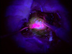
The trial, led by Colin Watts from the University of Cambridge, will treat patients with 5-Amino-Levulinic Acid (5-ALA), a metabolic marker of malignant glioma cells.  5-ALA is preferentially taken up by brain tumor cells and then converted into a strongly fluorescing porphyrin.

This conversion by the body of 5-ALA to a fluorescent chemical, shows the location of the glioblastoma when imaged under ultraviolet light.

Flourescent-5-ALA-Glioblastoma-Dr Colin Watts

The practical application of this is that it allows better identification of the tumor margins and avoids the removal of unnecessary brain tissue.

The use of fluorescent imaging to aid surgery is also being investigated in other tumors. Sally Church on Pharma Strategy Blog recently wrote about the use of folate receptor alpha fluorescence imaging in ovarian cancer.

It will be interesting to see how the GALA-5 phase 2 clinical trial in glioblastoma progresses.  The fluorescent imaging technology while promising is not without its pitfalls and potential risks.

As Jörg-Christian Tonn and Walter Stummer note in Vol 55 of “Clinical Neurosurgery (2008) some of the pitfalls with fluorescence-guided resection using ALA include:

  • Problem of overhanging margins i.e. inability to see all the tumor in the field of vision
  • Cysts leading to collapse of parts of the tumor with the result that areas are missed
  • Wrongly placed craniotomies preclude complete resection of contrast-enhancing tumor
  • Some cases of gliosarcoma show only modest fluorescence accumulation
  • Delay in drug administration may lead to less than optimal fluorescence
  • Exponential decrease in light with growing distance to fluorescent tissue, resulting in weak fluorescence intensity

No doubt many of the above issues will be controlled for in the GALA-5 trial. The challenge with imaging techniques is, however, in consistency and reproducibility outside of experienced clinical research investigation sites.

It will be interesting to see whether the potential pitfalls can be overcome such that this promising experimental imaging becomes routine in glioblastoma surgery.

Story Source: BBC Health

For those readers who would like to access Biotech Strategy Blog on their Kindle, this is a quick post to let you know it is now available on the Amazon Kindle Store for $0.99/month.  You can also continue to read it for free on the web.

Biotech-Strategy-Blog-Amazon-Kindle-StoreOf course, after Amazon takes its cut there is a (very) small royalty fee that ends up coming my way, so I have vested self-interest in promoting this.  However, it’s not something I anticipate getting rich from!

In a world of multi-channel marketing, it is good to try out new ideas that may make it easier for people to access content and information.

I grew up watching Lee Majors in the 1970’s TV show “The Six Million Dollar Man”, about an injured former astronaut whose bionic implants allowed him to do superhuman feats.

That was fiction, but it is becoming closer to reality as a result of new research into how artificial limbs can integrate with human tissues. This work on neural-electrical interfaces may ultimately allow someone to control a prosthesis as you would a normal limb, i.e. by nerve impulses that travel from the brain.

The promise of “functional integration” between the human nervous system and an electro-mechanical device is the greater control this would give amputees and potential for sensory feedback.

The laboratory of D. Kacy Cullen, Ph.D, assistant professor in the Department of Neurosurgery, Center for Brain Injury & Repair at the University of Pennsylvania, Perelman School of Medicine is actively working in this area. I had the pleasure to hear Dr Cullen talk about his research at Health Journalism 2011 earlier this year. I previously wrote on this blog about his research on nanomaterials that change color with blast impact.

Source: D Kacy Cullen PhD, University of Pennsylvania

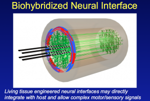
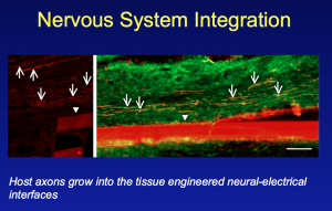
In his presentation to the Association of Health Care Journalists he described some of the neural tissue engineering work in his laboratory. This research has shown the ability to integrate axons in the peripheral nervous system (PNS) with an array of electrodes embedded in a living collagen matrix.

In essence this is the development of a nerve tissue/electrical interface that potentially allows the integration of man and machine.

Nervous System Integration


Key to success has been the development of a living, 3-D scaffold where this integration between nerve and electrodes can take place. Nerve axons in the peripheral nervous system require a living target for innervation. This has involved designing and engineering a 3-D living cellular matrix/scaffold that will work within a living body (to date the research has been on animal models).

Not only is Cullen and his laboratory looking at machine/nerve interfaces, but they have also developed techniques that may allow nerves to be repaired.  They have shown that nerves can be elongated or stretched using novel tissue engineering techniques.

The resulting axonal constructs via stretch-growth have been transplanted into rats and used to bridge an excised segment of sciatic nerve.  What was subsequently seen was an interwining plexus of host and graft axons, suggesting axonal regeneration across the lesion.

While still early stage, and not yet tested in humans, this research has tremendous potential for those paralysed due to traumatic nerve damage in the future.

Moving forward, the Penn researchers aim to develop an application for CNS tissue repair that can be delivered to the brain or spinal cord via stereotactic microinjection. This is minimally invasive surgery that allows delivery of cells to a precise location using 3D co-ordinates.

According to research published in Critical Reviews™ in Biomedical Engineering they plan to use micro-engineered hydrogel conduits, several centimeters long and the width of three hairs. These hydrogels will contain living axonal tracts that will then hopefully reconnect damaged nerves, and provide a platform or path for regeneration.

Many challenges still remain to be worked out for the tissue engineered neural constructs such as issues relating to inflammation and immune tolerance.

The research also needs to move from animals to humans, and be shown to be safe. The long-term functional outcome is also unknown.  It is far too early to think of this as a treatment option.

However, advances in neural tissue engineering, neuroregeneration and neuro-prosthetics do offer a lot of promise and hope to the many patients who suffer from spinal cord injuries or loss of a limb.

In a short blog post, I have not been able to do full justice to the innovative research or fully describe the techniques and methodology.  More information on the fascinating work being done at Penn, along with details of the associated scientific publications, can be found on the web page of the Cullen Laboratory: Neural Engineering in Neurotrauma.

ResearchBlogging.orgD. Kacy Cullen, John A. Wolf, Douglas H. Smith, & Bryan J. Pfister (2011). Neural Tissue Engineering for Neuroregeneration and Biohybridized Interface Microsystems In vivo (Part 2) Crit Rev Biomed Eng., 39 (3), 243-262


The patient advocacy session at the recent 16th Congress of the European Hematology Association in London focused on adherence to cancer treatments, and was filled to capacity, with the many attendees having to watch it from an overflow area.

Dr David Marin, Reader in Onco-Haematology at Imperial College, London presented research published last year in the Journal of Clinical Oncology that dramatically demonstrated how adherence to chronic myeloid leukemia (CML) therapy is the critical factor for achieving molecular responses.

In a study of 87 CML patients taking imatinib (Glivec®/Gleevec®) for a median period of 91 days, Dr Marin showed that no major molecular response (MMR) was observed when adherence was ≤ 80% and no complete molecular responses (CMR) were observed when adherence was ≤ 90%.  The graphical figure that he presented from his paper, dramatically shows how missing only a few doses of drug can have a major impact on outcome:

Source: Marin D, et al.  J Clin Oncol 2010; 28(14):2381-2388

Although the work by Marin and colleagues at the Hammersmith Hospital was undertaken with CML patients taking imatinib, the paper notes that adherence problems

“may apply equally to patients receiving second-generation tyrosine kinase inhibitors.”

Imatinib is the only TKI approved in the UK, thus that’s the only one available for studies there to date.

What made this data so compelling was the study rationale that used an electronic pill container, the medical event monitoring system (MEMS™) from the Aardex Group. This product contains a microchip that records the date and time it is opened.

Dr Marin’s study showed that “lack of adherence is underestimated by conventional methods.”  Self-reporting of adherence and pill counts are inaccurate compared to electronic data capture using MEMS (study subjects were unaware of the micro-chip in the pill bottle).
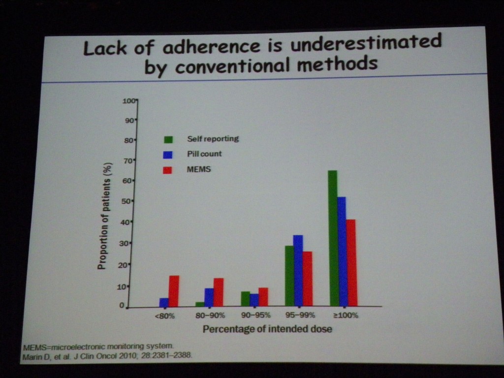
When psychologists at the Hammersmith Hospital subsequently interviewed patients who missed doses of drug, they found intentional and non-intentional adherence reasons.

A few excepts of  patient quotes from Dr Marin’s presentation:

Intentional non-adherence:

“Oh I can’t be bothered tonight, it’s not going to kill me [to miss a dose]”

“I thought there was no way I was going [on holiday] and being tired.”

Unintentional non-adherence:

“And sometimes you just forget”

“[the pharmacy] had no medication for me, so I went for nearly a week with no medication.”

Other speakers in the excellent patient advocacy session chaired by Jana Pelouchová (European Cancer Patient Coalition, Czech Republic) and Jan Geissler (CML Advocates Network, Germany) included Giora Sharf (Israeli CML patient’s Organization and CML Advocates Network, Israel) and Professor Rudolf Schoberberger (Medical University of Vienna, Austria).

Professor Schoberberger focused on the impact of drug packaging on compliance, particularly in elderly patients, and presented compelling research on how “child-proof” equals “age-proof.”  Sally Church in her video blog from EHA also discusses the patient advocacy session and how pharma/biotech companies could improve drug packaging.

The issue of adherence is a personal choice that every patient taking a chronic therapy makes. However, as Sally notes on Pharma Strategy Blog more patient and physician education is needed so that patients know there may be dramatic consequences from missing only a few doses per month.

Not only may adherence have a major impact on patient outcome, but as one questioner from France pointed out at the end of the EHA patient advocacy session, “for a statistician it is a nightmare.” Poor adherence in clinical trials “means that the true effect of a drug is not well known. Efficacy may be under-estimated if adherence is low.”

More monitoring of adherence in clinical trials through the use of MEMS technology may, therefore, be necessary to ensure that clinical trial data shows the true efficacy and adverse event profile of a drug.

I hope that the European Hematology Association (EHA) will make a webcast of this informative patient advocacy session publicly available online as it raised issues of considerable importance to patients, physicians and biotech/pharma companies alike.

ResearchBlogging.orgMarin, D., Bazeos, A., Mahon, F., Eliasson, L., Milojkovic, D., Bua, M., Apperley, J., Szydlo, R., Desai, R., Kozlowski, K., Paliompeis, C., Latham, V., Foroni, L., Molimard, M., Reid, A., Rezvani, K., de Lavallade, H., Guallar, C., Goldman, J., & Khorashad, J. (2010). Adherence Is the Critical Factor for Achieving Molecular Responses in Patients With Chronic Myeloid Leukemia Who Achieve Complete Cytogenetic Responses on Imatinib Journal of Clinical Oncology, 28 (14), 2381-2388 DOI: 10.1200/JCO.2009.26.3087
1 Comment

There is a lot of buzz this week about Lucentis versus Avastin for the treatment of wet age-related macular degeneration (AMD), something that will be talked about in more detail at the Association for Research in Vision and Ophthalmology (ARVO) annual meeting this weekend in Fort Lauderdale.

Also on the radar at ARVO is more news from Second Sight and their Argus II Retinal Prosthesis (something that I have previously written about on this blog).  For those interested there is a press conference at ARVO on Tuesday, May 3 from 5-6pm.

Second Sight presents updated results from the Argus II Retinal Prosthesis clinical trial, including sentence reading and color vision restoration for previously blind subjects. Two trial participants and independent investigators from the trial will be available for interviews.

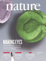
Which brings me back to a Nature article published earlier this month that I have been meaning to write about showing, for the first time, the ability to generate a three-dimensional culture of neural retinal tissue from mouse embryonic stem (ES) cells.  A word of warning, you may find the paper a little tough to follow unless you are a scientist in this field.

Eiraku and colleagues from Japan were able to culture retinal tissue similar to that seen in the human eye.  Eye formation starts as an optical vesicle that then develops into a two-walled optic cup.  As the authors note “optic cup development occurs in a complex environment affected by neighbouring tissues.”

What the authors showed in their research was the ability to culture retinal tissue containing ganglion cells, photoreceptors and bipolar cells.  They conclude:

Collectively, these findings demonstrate that the fully stratified neutral retina tissue architecture in this ES-cell culture self-forms in a spatiotemporally regulated manner mimicking in-vivo development.

My take on this research is that it is an important milestone in regenerative medicine that could lead to the prospect of retinal transplants in the future.  I look forward to learning more at ARVO about what the future may hold for retinal transplants derived from human stem cells.

ResearchBlogging.orgEiraku, M., Takata, N., Ishibashi, H., Kawada, M., Sakakura, E., Okuda, S., Sekiguchi, K., Adachi, T., & Sasai, Y. (2011). Self-organizing optic-cup morphogenesis in three-dimensional culture Nature, 472 (7341), 51-56 DOI: 10.1038/nature09941

The highlight of the recent Association of Health Care Journalists (AHCJ) annual meeting in Philadelphia (Health Journalism 2011) for me was the presentation by Kacy Cullen from the Center for Brain Injury and Repair in the Department of Neurosurgery at the University of Pennsylvania.

© Kacy Cullen, University of Pennsylvania

Dr Cullen presented his research on blast-induced traumatic brain injury (bTBI) and the development of a nanomaterial containing photonic crystals that change color upon exposure to blast pressure.

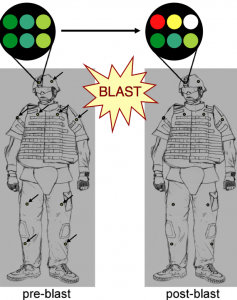
In the same way that a radiation dosimeter badge records exposure to cumulative radiation for a hospital worker, so a helmet-mounted color badge would change color based on a soldier’s exposure to blast pressure; a common occurrence with improvised explosive devices (IED).

In a paper published in NeuroImage, Cullen and colleagues describe in detail a blast-injury dosimeter (BID) made from photosensitive polymers that is like a colored sticker.  This nanomaterial contains microscopic, diamond-like photonic crystals, whose ability to refract light is damaged in a precise way by the pressure from explosive blasts.

The result is a change in color that is related to the degree of pressure and blast intensity. What’s more because the photonic crystals are structurally damaged by the blast, further exposure leads to more widespread microstructural alterations and a further change in color.  In essence, the crystals have a memory for cumulative blast exposure.

Why is this important?

Many soldiers are exposed to blasts, but show no overt symptoms of traumatic brain injury.  Research has shown that repeated hits to the helmet of a football player can lead to brain injury without the obvious signs of a concussion.  Traumatic brain injury as a result of repeated exposure to blasts may also lead to mild cognitive impairment and the possibility of increased risk for dementia, Alzheimer’s disease later in life.  This has been seen in NFL players.

The research by Cullen and colleagues is still in the early stages of development.  In their paper they acknowledge some of the next steps such as calibrating the color changes to levels of blast exposure, and correlating these with traumatic brain injury.  Any blast injury dosimeter will also need to be field tested.

However, this work is promising and an example of how nanotechnology may impact the detection and diagnosis of those soldiers at risk of traumatic brain injury.

War related scientific research often leads to civilian applications. In the future, I could see nanotechnology stickers that change color with cumulative impact on the helmets of NFL, college or high school football players.

You can read more about this innovative research on how color changing photonic crystals detect blast exposure in the journal NeuroImage.

Update June 30, 2011

If you are interested in the exciting and innovative research being undertaken by Kacy Cullen and his team, there is now a website for The Cullen Laboratory and their work on Neural Engineering in Neurotrauma.

ResearchBlogging.orgCullen, D., Xu, Y., Reneer, D., Browne, K., Geddes, J., Yang, S., & Smith, D. (2011). Color changing photonic crystals detect blast exposure NeuroImage, 54 DOI: 10.1016/j.neuroimage.2010.10.076

Today in the plenary session of the 102nd Annual Meeting of the American Association for Cancer Research (AACR), Lynda Chin from Dana-Farber Cancer Institute in Boston provided an excellent overview of the challenges and opportunities of translating insights from cancer genomics into personalized medicine that will benefit patients.

I unequivocally recommend listening to the webcast of the plenary when it is posted on the AACR website.

As Dr Chin stated at the start of her presentation, “cancer is fundamentally a disease of the genome.”  The goal of all cancer research is to make progress with prevention, detection and cure.

In the plenary presentation she highlighted some of the successes that have come from understanding the genome e.g. the knowledge of BRAF mutation in melanoma led to the identification of a target and development of a new drug in 8 years.  In addition to the development of novel therapeutics, genomics research has helped companies reposition drugs and she highlighted crizotinib as an example (move from C-Met to ALK inhibition in NSCLC).

These successes have “motivated researchers” according to Chin.  However, it is transformative new technology such as the next generation of sequencing technology that has heralded “a new era of cancer genomics.”  Massively parallel sequencing enables comprehensive genome characterization.

Not only has innovative new sequencing technology increased the throughput, but it has dramatically decreased the costs.  As Dr Chin noted, some have questioned whether cancer genomics is worth it?  She outlined some of the recent successes, such as BAP1 in ocular melanoma (see my previous post on this) as examples of its value.

Challenges remain such as the management of the vast amount of data that genome sequencing produces.  Data management, processing and storage remain issues, as does the need to develop a reference human genome against which a patient’s tumor profile could be compared.

And even when you find a mutation, the challenge is to separate the “drivers” from the “passengers.” This according to Chin requires a “robust statistical framework”.

Cancer signaling is not linear, but is a highly interconnected and redundant network, so it remains a big task to translate genomics into personalized medicine.  According to Dr Chin using mice as models to bridge the gap between sequencing and man may be the way forward in translating cancer genomics into personalized medicine.

Hospital marketing departments love new technology – the latest imaging, diagnostic or surgical equipment offers a point of differentiation from the competition.  This is particularly important in the United States where patients have a choice of hospital and surgeon.   Advertisements highlighting new technology are common, and patients actively seek out the “latest” option.

Today at the European Association of Urology (EAU) annual congress in Vienna, Associate Professsor Axel Merseburger from Hannover in Germany discussed some of the challenges with robotic surgery for prostatectomy or partial nepthrectomy.

  1. Lack of data showing an improved functional outcome compared to single port laparascopy or open surgery.  I was surprised that there are no comparative clinical trials that show robotic surgery to be better/worse than other surgical techniques. Complication rates remain inconclusive and urinary function is comparable. What is more, the panel of leading urologists concluded that high quality clinical trials would be difficult to design and enroll. One challenge in any surgical technique clinical trial is controlling for surgical experience; an important factor in determining outcome.   
  2. The need for licensing of robotic surgeons.  In the same way that airline pilots need to renew their licence every year and show they are competent in the skills required to fly a plane, there seemed to be concensus by the EAU panel that some form of “licensing” for robotic surgery should be required. However, as one member of the panel pointed out, it takes 250 patients to become proficient in new technology, which raises the issue of how that skill is obtained and if you were a patient, would you like to be one of those initial 250?
  3. The cost/benefit trade-off for robotic surgery remains unclear.  Robotic surgery takes longer, but is associated with shorter hospital stays, reduced blood loss and distinct cosmetic benefits. The fact that so much can be done through a small incision through the belly button is quite impressive.    However, the higher cost associated with the robotic procedures in terms of time, equipment and training has to be considered when there is no evidence of better functional outcome.  Do the benefits outweigh the costs?  The answer to that is not yet clear.

The take home that I took from the presentation by Dr Merseburger is that choice of surgeon is the key factor when facing any urology surgical procedure. As Dr Merseburger stated in one of his slides, “The risk of complication is related to the surgeons experience regardless of the surgical approach.” 

Those patients who are interested in robotic surgery should carefully consider the surgeon’s experience, with that particular equipment.  I expect we will see an ongoing debate about how innovations in surgical technology should be evaluated.

I would like to thank Victor Pikov, a neurophysiologist and biomedical engineer at Huntington Medical Research Institutes (HMRI) for drawing my attention to his NeurotechZone Blog that has a really fascinating post on the manufacturing of the next generation of artificial retina, the Argus™ 111, by the Lawrence Livermore National Laboratory (LLNL).

If you have an interest in this area, then Victor is also co-chair of 3rd International Conference on Neuroprosthetic Devices (ICNPD-2011) to be held in Sydney from November 25-26, 2011. Further information can be found on NeuroTechZone.

Second Sight Medical Products recently obtained a CE mark and European Market Approval for the Argus™ II system that incorporates 60 electrodes into the retinal prosthesis.

However the next generation of artificial retina, the Argus™ III is already in development.  It has 200 electrodes – a quantum leap forwards.  It’s hard not believe that an array that is four times as densely packed with sensors, will not provide improved vision.

Second Sight will no doubt be planning clinical trials for Argus™ III and it sounds like it will provide a further leap forward in the technology to restore some sense of vision to patients who have lost their sight through age-related macular degeneration (AMD) or retinitis pigmentosa (RP).

I have taken the liberty of embedding below, the excellent YouTube video that Lawrence Livermore National Laboratory (LLNL) have produced about their manufacturing of the Argus™ III artificial retina. It is well worth watching!


I wrote last week about Second Sight’s European Marketing Approval for the Argus II “artificial retina”.  What this news also stands for is the success of collaboration as a route to innovation.

The Artificial Retina Project (“Restoring Sight through Science”) through which Argus II was developed is a collaborative effort between six United States Department of Energy (DOE) research institutions, 4 universities and private industry.

Each offers unique scientific knowledge and specialist expertise, without which it is unlikely the project (that is continuing with the development of a more advanced Argus III artificial retina) would have been successful.

I’ve listed the collaborators below and as recorded on the DOE website, what they bring to the Artificial Retina Project.

DOE National Labs:

  • Argonne National Laboratory – Performs packaging and hermetic-seal research to protect the prosthetic device from the salty eye environment, using their R&D 100 award-winning ultrananocrystalline diamond technology.
  • Lawrence Livermore National Laboratory (LLNL) – Uses microfabrication technology to develop thin, flexible neural electrode arrays that conform to the retina’s curved shape. LLNL also uses advanced packaging technology and system-level integration to interconnect the electronics package and the thin-film electrode array.
  • Oak Ridge National Laboratory – Measures the effect of increasing the number of electrodes on the quality of the electrical signals used to stimulate the surviving neural cells in the retina.
  • Sandia National Laboratories – Develops microelectromechanical (MEMS) devices and high-voltage subsystems for advanced implant designs. These include microtools, electronics packaging, and application-specific integrated circuits (ASICs) to allow high-density interconnects and electrode arrays.
  • Brookhaven National Laboratory – Performs neuroscience imaging studies of the Model 1 retinal prosthesis.

Universities:

  • Doheny Eye Institute at the University of Southern California – Provides medical direction and performs preclinical and clinical testing of the electrode array implants. Leads the Artificial Retina Project.
  • University of California, Santa Cruz – Performs bidirectional telemetry for wireless communication and chip design for stimulating the electrode array.
  • North Carolina State University – Performs electromagnetic and thermal modeling of the device to help determine how much energy can be used to stimulate the remaining nondiseased cells.
  • California Institute of Technology – Performs real-time image processing of miniature camera output and provides optimization of visual perception.

In October 2004, Second Sight Medical Products and the DOE signed a Co-Operative Research and Development Agreement (CRADA) in which the above institutions agreed to share intellectual property and royalties from their research, with Second Sight chosen to be the commercial partner.  As part of the CRADA, Second Sight obtained a limited, exclusive license to the inventions developed during the DOE Retinal Prosthesis Project.

You can find more information about the history of this fascinating project on the Artificial Retina Project website, that also has links to several patient stories from around the world.

The Artificial Retina Project is a case study on the success of collaboration.  Whether such an ambitious project that was funded by the US Government would ever have taken place in the private sector is the question that comes to my mind?  Would a private company have been able to harness the intellectual power of 10 research institutions in this way?

If not, then do governments have a role to play in biomedical innovation by drawing partners together so that advances in basic research can be applied to new products, whether they be new drugs or novel devices?

And if you agree that governments do have a role to play what should be the extent of government funding?  In the case of artificial retina, the DOE has funded this since 1999, with its contribution rising from $500K to $7M per year. Those numbers may also be direct costs, and not reflect the cost of investments in buildings, research facilities etc.

I’d be interested in any thoughts you would like to share on this.

error: Content is protected !!