Biotech Strategy Blog

Commentary on Science, Innovation & New Products with a focus on Oncology, Hematology & Immunotherapy

About Pieter Droppert

Here are my most recent posts

Posts by Pieter Droppert

At the 2012 European Society for Medical Oncology (ESMO) meeting in Vienna today, the first published clinical data for a new second generation anti-androgen (ODM-201) was presented. Company representatives inform me that the poster will be available on the Orion Pharma website in a few days. (Update Oct 9: it is now available, but all the text on the PDF of the poster available for download appears to have been intentionally blurred to make it unreadable!)

What makes ODM-201 interesting? The company claimed on their poster, “it is a uniquely designed AR antagonist” yet citing confidentiality reasons refused to answer questions about it or offer a comparison of their drug to other second-generation androgen receptor (AR) antagonists ahead of them in development such as ARN-509, or enzalutamide (Xtandi), which was recently approved in the United States. This perhaps reflects their inexperience as an oncology drug development company, and was a missed PR opportunity.

What the Orion Pharma poster does say is that ODM-201 has “negligble brain penetrance” in nonclinical models. If there are CNS problems with enzalutamide then this would be a major potential advantage, but a more important question in my view is whether ODM-201 has activity against splice variants? The company declined to answer.

Joan Carles, MD PhD (Vall d’Hebron, Barcelona) was the poster discussant and summed up the phase 1 dose escalation trial with 18 patients by summarizing the strengths and weaknesses of the data presented for ODM-201:

Strengths

  • High efficacy
  • Well tolerated
  • Linear pharmacokinetics

Weakness

  • Few patients

We will have to wait for more trial results with ODM-201 and more information on its mechanism of action before it’s potential can be properly evaluated, but at first glance it appears to offer promise.

However, Orion Pharma are seeking to enter a very busy and competitive prostate cancer market. Some of the challenges it faces will be:

  • Which agent to use as a comparator when it moves into randomized trials?  The days of placebo controlled trials in advanced prostate cancer appear to be now over, and most likely it would have to compare itself against enzalutamide if this is the standard of care it will be competing against.
  • Will it need to combine with other agents to be successful? The future is now moving towards combinations. Presuming ODM-201 continues to show promise, will Orion Pharma combine ODM-201 with another novel agent e.g. a PI3K inhibitor? Will they have the courage to do a novel-novel phase 2 combination trial?

The prostate cancer landscape just got even busier with the arrival of ODM-201 on the scene and it will be interesting to watch the drug development strategy of Orion Pharma in partnership with Endo Pharmaceuticals, who have a pain and urology franchise.

I certainly look forward to seeing more data on ODM-201 at future medical meetings and evaluating it against enzalutamide as more data becomes available.

At the 2012 Congress of the European Society for Medical Oncology (ESMO 2012) in Vienna,  Mitesh J. Borad MD, Assistant Professor of Medicine at the Mayo Clinic in Scottsdale, AZ presented the results of the TH-CR-404 phase 2 clinical trial that compared the efficacy and safety of TH-302 (Threshold Pharmaceuticals) plus gemicitabine versus gemcitabine alone in patients with untreated advanced pancreatic cancer.

TH-302 is an experimental cancer drug in development that kills cells i.e. is cytotoxic under conditions of low oxygen (hypoxia) found in the microenvironment of cancer tumors. Sally Church, PhD on Pharma Strategy Blog has written about the mechanism of action for TH-302 and the results presented earlier this year at the 2012 annual meeting of the American Association for Cancer Research (AACR).

Subscribers to Premium Content can login to read about the ESMO 2012 results and why the results fail to impress.

This content is restricted to subscribers

In the phase IIb study presented at AACR 2012, TH-302 at a dose of 240mg/m² showed a progression free survival benefit of 2 months over gemcitabine. I encourage you to read her article.

ESMO 2012 Results

At ESMO 2012 in the Vienna, the TH-CR-404 data presented (abstract 6660 – full presentation available on the Threshold Pharmaceutials site) showed a benefit of 2.4 months in median progression free survival (PFS) for those patients receiving TH-302 at a dose of 340mg/m2 + gemcitabine compared to gemcitabine alone.

The PFS was only significant with the 340mg/m² dose and overall survival (OS) was not significant for either dose. However it should be noted that OS was not a primary endpoint for the study, nor was it powered to detect OS.

Hematological Toxicities may be difficult to handle

The 2.4 month (median) increase in PFS seen with TH-302 + gemcitabine does come with increased toxicities compared to gemcitabine alone.  These include an increase in Grade 3 or 4 thrombocytopenia (63%), and increase in Grade 3 or 4 neutropenia (60%). As a UK oncologist sitting next to me said after the presentation, “it is interesting, but quite toxic.”

Threshold plan to start a Phase 3 Trial

Dr Borad told the ESMO 2012 audience that Theshold planned to initiate a randomized phase III trial of TH-302 in advanced pancreatic cancer with the 340mg/m² dose.

Presentation Discussion at ESMO 2012

Michel Ducreux, Head of the Institut Gustave Roussy, Villejuif, France and Professor of Medicine at the University of Paris Sud was the discussant and put the data in context for the audience:

Some of the key points he made were:

  • A drug that targets hypoxia may be interesting in pancreatic cancer.
  • It was a well done, randomized phase 2 study
  • Concern about high level of hematological toxicity.

Professor Ducreux told the audience the hematological toxicity of TH-302 + gemcitabine would be difficult to handle in the many small hospitals in France if it becomes a standard of care. For these reasons, it also means such a combination may be disliked by community oncologists in the United States.  Similarly, FOLFIRINOX has shown efficacy versus gemcitabine, but more widespread uptake in first line has been limited by the severe toxicities (including chronic diarrhea) induced by the combination and is therefore not well liked by many oncologists.

“The results are not too bad” said Ducreux. However, he went on to to say “we have to remain suspicious” because positive phase 2 results can be followed by a totally negative randomized phase III trial. He gave axitinib as an example.

“The treatment of pancreatic cancer remains very difficult and I am happy to see new results, but again we have to stay on evidence based medicine, and it is good a phase 3 is planned to evaluate this new compound,” said Ducreux.

Why the results fail to impress

The ESMO 2012 data for TH-302 does, however, have to be considered in the light of the fact that there are other on-going trials that could change the standard of care.

One of trials highlighted by Ducreux in his discussion was Celgene’s nab-paclitaxel (Abraxane) phase III trial that I predict will have positive data in the near future. Here’s a link to my Storify from ESMO 2012 on Abraxane that discusses this.

Professor Ducruex told the ESMO 2012 audience that we will have the results for nab-paclitaxel probably for the next ASCO GI.

If nab-paclitaxel were to increase overall survival to 10-12 months, this would become the new standard of care that TH-302 would then have to beat.  Based on the TH-302 data presented at ESMO 2012, even if you ignore the hematological toxicity, this is hard to imagine from the phase II study, which is why the data failed to impress me.

However, until we have the TH-302 phase III data we will not know for sure. In the meantime I am looking forward to the possibility of the Abraxane pancreatic data at ASCO GI and a major breakthrough in the treatment pancreatic cancer. The prospect of this happening will offer hope to all those patients with pancreatic cancer, in a disease that for so long has been a graveyard of drug development.


1 Comment

One of the late-breaking abstracts (not yet published) that I am looking forward to at the forthcoming annual Congress of the European Society for Medical Oncology (ESMO 2012) in Vienna is on ODM-201 (Orion Pharma):

LBA25-PR:  ARADES trial: A first-in-man, open-label, phase I/II safety, pharmacokinetic, and proof-of-concept study of ODM-201 in patients (pts) with progressive metastatic castration-resistant prostate cancer (mCRPC)

ODM-201 is a new antiandrogen from Finnish company, Orion Pharma, and is being developed in partnership with Endo Pharmaceuticals (NASDAQ: ENDP).

This content is restricted to subscribers

In a corporate presentation, Orion Pharma describe ODM-201 as:

  • Potentially best-in-class antiandrogen
  • Does not enter brain in preclinical models
  • No testosterone increase in animal models
  • Well tolerated

“We are studying and developing an anti-androgen with qualities that currently cannot be found in any of our or our competitors’ drugs”

says Mika Mustonen (@MikaMustonen), Head of Oncology, Research and Development at Orion in an article, “Pursuing a targeted drug for prostate cancer” published by the company. Mustonen says:

“The research on our new drug candidate, ODM-201, suggests that we may be able to provide patients with a new alternative for the treatment of prostate cancer.”

Of note, is Orion’s focus on biomarkers, which may help predict which patients are more likely to respond to the therapy. According to Mustonen:

“Biomarkers increase the chance of success.  By following them we can study topics that have not been considered before in this type of research.  We can predict different phases of the disease, survey any safety risks associated with the drug and find out what kind of patients benefit most from the drug.”

At ESMO 2012 (Twitter hashtag #ESMO12) I expect we will hear preliminary data from the ARADES 3104001 phase 1 dose escalation study (NCT01317641) with ODM-201.

According to clinicaltrials.gov this multicenter, non-randomized clinical trial is being undertaken at sites in Finland, Czech Republic, France, United Kingdom and the United States.

After 12 weeks in the phase 1 dose escalation study, patients with stable disease can continue treatment in a phase 2 extension study on the safety and tolerability of ODM-201 (NCT01429064).

As of May 2012, Orion Pharma reported that the ARADES 3104001 phase II expansion component had 105 patients enrolled, with 3 dose levels to be expanded.

Company senior management have told me they “are very excited about the ODM-201 data,” to be presented at ESMO. I have not seen the data, but presume the results will be positive. After all, company executives don’t get excited about negative data!

Is there a market for a new antiandrogen?

Although Medivation are first to market with their androgen receptor (AR) inhibitor, enzalutamide/MDV3100 (Xtandi) that does not mean that other companies will not be able to make in-roads into the market with cheaper or more effective AR antagonists.

In a Pharma Strategy Blog interview with Sally Church, Dr Charles Sawyers noted that Aragon’s ARN-509 (another AR inhibitor in development) is “more potent” than enzalutamide and “might produce a higher percentage of responders or longer duration of response.”

Medivation recently announced that enzalatumide is available in the United States for patients with metastatic castration resistant prostate cancer previously treated with docetaxel.

At a price of $7,450 a month, however, Xtandi is considerably higher than Johnson & Johnson’s Zytiga.  This aggressive premium pricing strategy opens the door to competitors who may offer equally effective, but less expensive drugs.

The prostate cancer market remains a dynamic one and very much one to watch over the next few years.

I look forward to learning more about ODM-201 at ESMO 2012.

6 Comments

Can you imagine what it must be like to go blind? Degenerative diseases of the eye such as age-related macular degeneration (AMD) and retinitis pigmentosa (RP) can result in loss of vision and blindness.

Retinitis pigmentosa is an inherited genetic condition for which there is no cure. It results in the progressive loss of function of the retinal photoreceptors that convert light into electrical nerve impulses that travel down the optic nerve to the brain for processing into the images we see. As you reduce the ability to process light, so you start to lose your sight and can end up totally blind.

Several companies and research groups are now working on an artificial retinal prosthesis. The term “bionic eye” is often used to describe this research, which for many may conjure up the “$6 million dollar man“. This “hype” is, however, far from the reality of where the technology currently is at.

To date, only crude images can be detected. There is no dramatic restoration of sight or vision, instead the retinal prostheses currently only offer the ability to detect light and large shapes.

However, for those who are blind, being able to detect shapes and “see” a door, could help people enormously in their activities of daily living. In the same way that we have seen tremendous increase in the megapixels for digital cameras, so it is hoped that visual acuity will improve as the technology increases the number of pixels that can be processed.

There are several research groups and companies working on different types of electrodes and different implant locations in the retina, but in essence they have a common approach.

Most of the retinal prostheses in development use an external video camera to convert light to electrical signals that are transmitted wirelessly to a retinal implant (multielectrode array) that delivers a pattern of electrical signals to the retina. This electrical stimulation of the retinal cells results in signals being passed down the optic nerve to the brain where they are processed into visual images.

The Argus II Retinal Prosthesis System (Second Sight) is the most advanced in terms of commercial approval. It has obtained CE marking and is on the market in Europe where it sells for a price of $115,000 (Source: ExtremeTech).

On September 28, 2012, the FDA ophthalmics device panel will discuss the application by Second Sight to sell the Argus II system in the United States as a humanitarian use device.

The Argus II implant consists of a coil (for receiving and transmitting wireless data) fixed on the sclera (outside of eye) and a 60 electrode array positioned on the surface of the retina.

Argus II Clinical Trial Results

The results of a multicenter trial with 30 patients at 10 centers were published earlier this year in Ophthalmology, a journal of the American Academy of Ophthalmology (AAO). Mark S. Humayun from the Doheny Eye Institute at University of Southern California and research colleagues reported that:

“Subjects performed statistically better with the system on versus off in the following tasks: object localization (96% of subjects), motion discrimination (57%), and discrimination of oriented gratings (23%). The best recorded visual acuity to date is 20/1260.”

In addition to finding the device to be reliable over the long-term, Humayun et al concluded that:

“The data in this report suggest that, on average, prosthesis subjects have improved visual acuity from light perception to at least hand movements, with some improving to at least counting fingers.”

“These visual acuity data and other performance results to date…demonstrate the ability of this retinal implant to provide meaningful visual perception and usefulness to subjects blind as a result of end-stage outer retinal degenerations.”

Bionic Vision Australia announces first implant of Bionic Eye

Last month, Bionic Vision Australia, announced in an August 30, 2012 media release that they had “successfully performed the first implantation of an early prototype bionic eye with 24 electrodes.”

The recipient, Dianne Ashworth is quoted as saying:

 “I didn’t know what to expect, but all of a sudden, I could see a little flash…it was amazing. Every time there was stimulation there was a different shape that appeared in front of my eye,”

Veronika Gouskova, Marketing and Communications Manager of Bionic Vision Australia (BVA) kindly responded by email to my questions about their news:

BSB: I saw in your Aug 30 media release that the implant was described as a “world first” by Dr Allen. Could you clarify in what way this is a “world first” when there are other retinal prosthetics on the market e.g. Argus II from Second Sight?

BVA: This is a world first implantation of a device in the suprachoroidal space at the back of the eye. Although there have been other groups working with patients around the world, this surgical position and procedure is unique to Bionic Vision Australia

BSB: How does the Australian Bionic Eye differ from the Second Sight product that is marketed in Europe?

BVA: There are a number of groups internationally working in the field of retinal implants – the fact that this research is ongoing means that the problem hasn’t yet been solved completely. Between all these groups there are a differences in technology being developed, materials used, surgical placement of the device, surgical technique and the way the electrodes or photodiodes are being stimulated (i.e. how the visual data is processed before it is sent to the implant).

At the end of the day, it’s all about ensuring the best outcome for patients. We are developing two prototypes, with different functional aims: the ‘wide-view’ device combines novel technologies with materials that have been successfully used in other clinical implants. This approach incorporates a microchip with 98 stimulating electrodes and aims to provide increased mobility for patients to help them move safely in their environment.

Our ‘high-acuity’ device incorporates a number of exciting and new technologies, such as diamond materials,  to bring together a microchip and an implant with 1024 electrodes. The device aims to provide functional central vision to assist with tasks such as face recognition and reading large print. The early prototype that we implanted with our first three patients is a stepping stone towards further development for these two devices.

Bionic Vision Australia brings together researchers from many different fields, so we have a truly multidisciplinary team. This means the clinicians and surgeons are involved in the design and development process from the start. Further, a lot of our researchers were involved in the cochlear implant, or bionic ear development – they know what it takes to bring a competitive medical implant to the market.

BSB: I saw that your device was placed between the choroid and sclera, is that significant as opposed to being on the top of the retina? Could you provide further clarification on the significance of this and the surgical technique required for implantation?

BVA: Yes, this early prototype is implanted in the suprachoroidal space, between the choroid and the sclera. This is beneath the retina. There are a number of advantages in doing this, e.g. this position greatly enhances the mechanical stability of the implant and allows for a relatively straightforward surgery. The surgical procedure involves making an incision through the sclera and sliding the implant in place.

BSB: What are the next steps, is a larger clinical trial of your device already planned/underway – when will other patients receive implants?

BVA: We have implanted this early prototype in three patients and will continue to work with these patients over the next 18 months while we further develop our full prototype devices. The next step will be of course, a larger trial with our full devices in due course.

BSB: How might your device be potentially sold/commercialized or made available – is there a commercial partner associated with Bionic Vision Australia?

BVA: Bionic Vision Australia is an unincorporated joint venture between a number of research organisations in Australia. We are funded by the Australian Research Council. A commercialisation vehicle has been set up, Bionic Vision Technologies, to commercialise the technology. This company is solely owned by the member organisations that are involved in our research.

In addition to Second Sight and Bionic Vision Australia, there are several other companies and research groups with retinal prostheses in development.

It is an area where we are seeing innovation in action as teams of multi-disciplinary researchers strive to restore sight and improve the quality of life to people who have become blind.

However, given the high cost of R&D, and the fact that much of the research is government funded, I’m not sure of the commercial opportunity. It will be interesting to see how the market for retinal prostheses develops.

Challenges that have to be overcome

A number of challenges with retinal prostheses will have to be overcome. Some of these are discussed in a 2011 editorial by James Weiland, Alice Cho and Mark Humayun on “Retinal prostheses: Current Clinical Results and Future Needs” that was published in the AAO journal, “Ophthalmology.”  I encourage anyone with an interest in this area to read this insightful review.

Some of the questions I took from this editorial were:

  • Why do some patients respond better than others with an implant?
  • Will retinal remodeling and ongoing degeneration limit the usefulness of implants?
  • What is the best surgical way to ensure optimal placement of the stimulating arrays to maximize visual acuity but avoid problems associated with fixation, and wound closure?
  • Could artificial vision be detrimental to other sensory inputs e.g. the ability of the visual cortex to process data from non-visual Braille reading?

Answers to these and other questions will come as clinical experience is gained and advances in technology improve the design and functionality. In my view this is innovation in action.

Update February 15, 2013 Argus II receives FDA approval

As expected following the positive endorsement of the Ophthalmic Devices Advisory Panel last year, the United States Food and Drug Administration (FDA) announced in a Feb 14, 2013 press release that Second Sight’s Argus II retinal prosthesis system had received approval as a humanitarian use device for the treatment of adults with advanced retinitis pigmentosa.


References

ResearchBlogging.orgHumayun MS, Dorn JD, da Cruz L, Dagnelie G, Sahel JA, Stanga PE, Cideciyan AV, Duncan JL, Eliott D, Filley E, Ho AC, Santos A, Safran AB, Arditi A, Del Priore LV, Greenberg RJ, & Argus II Study Group (2012). Interim results from the international trial of Second Sight’s visual prosthesis. Ophthalmology, 119 (4), 779-88 PMID: 22244176

Weiland JD, Cho AK, & Humayun MS (2011). Retinal prostheses: current clinical results and future needs. Ophthalmology, 118 (11), 2227-37 PMID: 22047893

1 Comment
Drug development for neurodegenerative brain diseases such as Parkinson’s or dementia, of which Alzheimer’s is the most common form, needs to focus on patients early in the disease, not those where brain damage has already occurred.

Diagnosing and treating patients more effectively earlier will, even if you aren’t able to instigate a cure, offer the ability to modify the disease progression and slow or delay when brain damage occurs.  In the case of Alzheimer’s, once the amyloid plaques (tangles of misshapen proteins) have accumulated in nervous tissue, it has so far been impossible to untangle or remove them.

Subscribers can login to read more below:

This content is restricted to subscribers

Last year, I interviewed Dr Todd Sherer, (then the Chief Program Officer) and now the CEO of the Michael J. Fox Foundation, who told me that: “biomarkers are a real focus of the foundation.” Sherer went on to say that:

“Parkinson’s is a difficult disease to diagnose, there is no definitive diagnostic test, so it ends up a clinical diagnosis.  Getting a biomarker that could help better confirm the diagnosis would allow people to get the correct treatment earlier in their disease”

Which is why I was interested to see new research published earlier this week in the journal Archives of Neurology (online first, August 27, 2012), by Sara Hall and colleagues at Lund University, University of Gothenburg and Skåne University Hospital in Malmo, Sweden.

Hall and colleagues describe how a panel of five cerebrospinal fluid (CSF) biomarkers allowed the differential diagnosis of common dementia from Parkinsonian disorders:

  • Beta-amyloid 42
  • Total tau
  • Phosphorylated tau
  • Alpha-synuclein
  • Neurofilament light chain

Patients with early symptoms of neurodegenerative diseases can be hard to diagnose.  Misdiagnosis can occur, which means patients may not respond to treatment or they could be enrolled into a clinical trial, and end up skewing the results.

Ensuring that we have the right patients in clinical trials is important as we seek to alter disease progression.  In other words it’s important to see whether new drugs or treatments are impacting the disease course.  If you have a wrongly diagnosed patient in a trial, then the drug may show no effect, not because it’s not effective, but that patient’s disease is not responsive.

Multivariate analysis indicated that the panel of 5 CSF biomarkers could accurately differentiate Alzheimer’s disease (AD) from Parkinson disease with dementia (PDD), and dementia with Lewy bodies (DLB). The Neurofilament light chain biomarker alone could differentiate PD from atypical Parkinson disease, Hall and colleagues noted.

Whilst the panel was not able to distinguish all forms of dementia, in an accompanying editorial Richard J. Perrin MD, PhD from the University of Washington, stated that this research “represents a significant step forward.” Perrin concluded that:

“Implementation of CSF biomarker panels such as this one should improve the efficiency of clinical trials and accelerate the evaluation and discovery of new effective treatments for neurological diseases.”

Summary

Developing biomarkers that assist in the ability to diagnose Alzheimer’s, Parkinson and dementia patients correctly, and then be able to monitor their subsequent disease progression, should be a key focus of those biotechnology and pharmaceutical companies that want to do innovative and rational drug development.

References

ResearchBlogging.orgSara Hall, MD, Annika Ohrfelt, PhD, Radu Constantinescu, MD, Ulf Andreasson, PhD, Yulia Surova, MD, Fredrik Bostrom, MD, Christer Nilsson, MD, PhD, Hakan Widner, MD, PhD, Hilde Decraemer, Katarina Nagga, MD, PhD, Lennart Minthon, MD, PhD, Elisabet Londos, MD, PhD, Eugeen Vanmechelen, PhD, Bjorn Holmberg, MD, PhD, Henrik Zetterberg, MD, PhD, Kaj Blennow, MD, PhD, & Oskar Hansson, MD, PhD (2012). Accuracy of a Panel of 5 Cerebrospinal Fluid Biomarkers in the Differential Diagnosis of Patients With Dementia and/or Parkinsonian Disorders Arch Neurol. DOI: 10.1001/archneurol.2012.1654

Richard J. Perrin, MD, PhD (2012). Cerebrospinal Fluid Biomarkers for Clinical Trials Arch Neurol. (August 27 Online First) DOI: 10.1001/archneurol.2012.2353

What is a Biomarker?

According to the Biomarkers Definitions Working Group, a biomarker is:

“a characteristic that is objectively measured and evaluated as an indicator of normal biologic processes, pathogenic processes, or pharmacologic responses to a therapeutic intervention.”

An example of a common biomarker is blood pressure. High blood pressure is a surrogate for cardiovascular disease and risk of stroke.

Why are biomarkers important? Subscribers can login to read more below.

This content is restricted to subscribers

Why are Biomarkers important?

Biomarkers can be used for diagnosis and for monitoring the safety and effectiveness of treatments. They are increasingly becoming important in the selection of patients for clinical trials, and as potential surrogates for clinical endpoints that may take a long time to occur e.g. measuring how long someone will live in a cancer trial (overall survival).

Examples of the use of biomarkers include:

  • Diagnosis: high blood pressure is used as a biomarker for cardiovascular disease and risk of stroke.
  • Treatment Selection: CSF biomarkers that correlate with neurodegenerative diseases may help select the most appropriate treatment
  • Drug Effectiveness: biomarkers can be used to monitor treatment or drug effectiveness e.g. use of cholesterol levels as a measure of cardiovascular disease
  • Surrogate Clinical Endpoint: a biomarker based on scientific evidence that predicts or correlates with clinical benefit could be used as a surrogate for a clinical endpoint that may take a while to detect e.g. how long a patient lives or survives, and in the process speed up drug development. Recent prostate cancer trials sought to show that circulating tumor cell (CTC) counts correlated with the survival benefits seen. However, validation of a biomarker needs to take place before regulatory agencies will accept it as a surrogate endpoint in clinical trials.

Biomarkers can be divided into those which are prognostic and those that are predictive.

Prognostic Biomarker: a marker that provides information on the likely course of a disease in an untreated individual.

Prognostic biomarkers are used to identify high-risk cancer patients who should, therefore, receive adjuvant therapy.

Predictive Biomarker: a marker that provides information on how likely you are to respond to a particular therapy.

Predictive biomarkers are used to guide treatment choices i.e. selecting the therapy with the highest likelihood of success.

In breast cancer, estrogen and progesterone receptors are biomarkers that predict sensitivity to endocrine therapy, while HER2 levels predict response to Herceptin treatment. In colorectal cancer (CRC) patients, KRAS mutations have been shown to be a biomarker of resistance to EGFR targeting drugs such as cetuximab and panitumumab.

Predictive biomarkers allow expensive new cancer treatments to be given only to those patients who are likely to respond. As we move forward into the era of personalized medicine the aim is to develop more highly predictive biomarkers that will allow better detection, diagnosis and treatment of disease.

In addition, there’s also a need to develop biomarkers that can distinguish between subgroups of patients to separate those who might benefit from a therapy and those who have developed resistance. Biomarkers for resistance to cancer therapy is an increasingly important area of research.

For those readers interested in cancer biomarkers, the joint ASCO-EORTC-NCI “Markers in Cancer” 2012 meeting in Hollywood, FL (near Fort Lauderdale) from October 11-13 has an agenda that holds promise.

Some of the presentations that caught my attention and ones I particularly look forward to watching remotely via the “Virtual Meeting” include:

  • Biomarkers of Resistance to EGFR-Targeted Therapies in Lung Cancer
    Enriqueta Felip, MD, PhD – Vall d’Hebron University Hospital
  • Resistance Mechanisms to BRAF Inhibition in Melanoma
    Jeffrey Sosman, MD – Vanderbilt-Ingram Cancer Center
  • Complexities of Identifying Non-Mutational Biomarkers of Resistance:
    The VEGF Pathway Example
    Michael B. Atkins, MD – Georgetown University
  • Development of Biomarkers for PI3K Pathway Targeting
    Sherene Loi, MD, PhD – Jules Bordet Institute, Brussels
  • Emerging Functional Imaging Biomarkers
    Annick D. Van Den Abbeele, MD – Dana-Farber Cancer Institute

The next post in this mini-series will discuss new research that shows how a panel of 5 CSF biomarkers can be used to differentiate between neurodegenerative diseases that might otherwise be misdiagnosed. This is particularly important for clinical trial recruitment where early symptomatic patients could potentially be recruited in error if given the wrong diagnosis, and placed in trials that they will not respond to.

Regular blog readers will know I think tissue engineering is an exciting area where you can see innovation in action – advances in basic science can translate into ways to artificially create replacement organs and body parts.

Research published online 22 July 2012 in Nature Biotechnology by Janna Nawroth and colleagues at the California Institute of Technology (Caltech) and Harvard University, shows how biomedical engineers are learning from the structure and function of other animals.

In a Nature Biotechnology article titled “a tissue-engineered jellyfish with biomimetic propulsion” researchers describe how they were able to combine rat cardiac muscle cells and a synthetic elastomer membrane into a medusoid like structure that mimicked the propulsion of a jellyfish.

Credit: Caltech and Harvard University

They achieved this by forming the elastomer into a medusoid or jellyfish like shape with eight lobes around a central disc, then applying a monolayer of rat cardiac muscle tissue, which when electrically shocked, contracted in a synchronized way.

The net result was the medusoid “swam” in a similar way to a jellyfish. They effectively developed an artificial pump made out of a hybrid of living cells and silicone rubber.

The video below by Janna Nawroth, produced by Caltech and Harvard University, shows the medusoid in action, and explains how this research advances the design of muscular pumps for biomedical application:

According to the Caltech press release, this approach in reverse-engineering the function of a jellyfish “will be broadly applicable to the reverse engineering of muscular organs in humans.” 

While we are not yet able to tissue engineer a replacement human heart, it’s hard not to believe that at some point in the future we will see the development of hybrid devices that combine synthetic materials and cultured heart muscle cells.

Reference

ResearchBlogging.orgJanna C Nawroth, Hyungsuk Lee, Adam W Feinberg, Crystal M Ripplinger, Megan L McCain, Anna Grosberg, John O Dabiri, & Kevin Kit Parker (2012). A tissue-engineered jellyfish with biomimetic propulsion Nature Biotechnology, 30, 792-797 DOI: 10.1038/nbt.2269

Imagine that you are born deaf and live in a world of silence – what price would you pay for a new treatment that might restore your hearing?
That is the market opportunity that may be available for biotechnology and pharmaceutical companies as the basic science around congenital hearing loss starts to yield insights that could translate into new products.

Research published in the July 26, 2012 issue of the journal “Neuron” by Omar Akil from UCSF and colleagues at the University of Pittsburgh and Ohio State University, showed the ability to reverse hearing loss in mice through the use of gene therapy (viral-mediated insertion) to replace the absent vesicular glutamate transporter-3 gene (VGLUT3).

VGLUT3 is a gene involved with the transport of the neurotransmitter glutamate that is required by inner hair cells in order to generate neural responses to sound. Mice lacking VGLUT3 can’t hear.

Insertion of the VGLUT3 gene into mice cochlear cells resulted in restoration of hearing that lasted for 9 months (that’s a long time for mice). The authors noted that:

“These findings represent a successful restoration of hearing by gene replacement in mice, which is a significant advance toward gene therapy of human deafness.”

Over 50% of all human hearing loss is genetically based, and as tools to understand the human genome develop, scientists have been able to identify a number of genes associated with hearing loss.

Research in animal models is ongoing, with the potential in the future that we may be able to replace, repair or correct a defect a genetic mutation.

Could this lead to the restoration of human hearing? The answer is “yes”.

An accompanying editorial in Neuron by Donna Martin and Yehoash Raphael from The University of Michigan describes the work by Akil and colleagues as a major breakthrough:

“Results presented in their paper are a true breakthrough because they show that gene therapy can lead to functional recovery from sensorineural deafness. Even more exciting is the direct relevance of this work to a large population of humans who have mutations in the VGLUT3 gene.”

There remain a number of challenges before gene therapy to correct human deafness becomes a reality, but biopharmaceutical companies such as GenVec (NASDAQ: GNVC) already see the market opportunity and potential for gene therapy to correct hearing loss. Novartis have a collaboration agreement with GenVec that is worth up to $213.6M in milestone payments.

The potential of gene therapy to restore hearing loss will offer hope to many with deafness. It is an exciting area to watch as innovative science translates into personalized medicine.

References

ResearchBlogging.orgOmar Akil, Rebecca P. Seal, Kevin Burke, Chuansong Wang, Aurash Alemi, Matthew During, Robert H. Edwards, & Lawrence R. Lustig (2012). Restoration of Hearing in the VGLUT3 Knockout Mouse Using Virally Mediated Gene Therapy Neuron, 283-293 DOI: 10.1016/j.neuron.2012.05.019

Donna M. Martin, & Yehoash Raphael (2012). Have You Heard? Viral-Mediated Gene Therapy Restores Hearing Neuron, 75, 188-190 DOI: 10.1016/j.neuron.2012.06.008

5 Comments

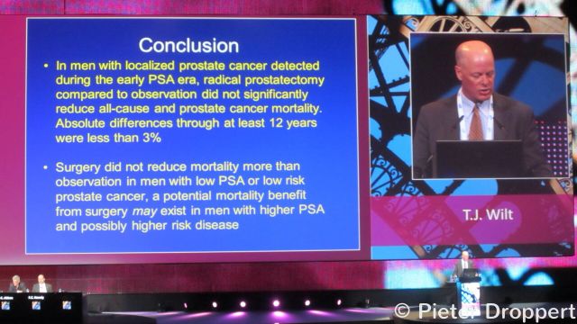
Dr Benjamin J. Davies, an academic urologist at the University of Pittsburgh today castrated the media over their coverage of the Prostate Cancer Intervention versus Observation Trial (PIVOT).

Pivot Prostate Cancer Trial Conclusion

In an article titled “Prostate Cancer: Lessons from PIVOT lost in media hype” published in the News and Views section of Nature Reviews Urology, Dr Davies states, “we must be careful to ensure the less-newsworthy facts and limitations of high-profile trials, such as PIVOT, are not lost in the media hype.

Davies goes on to say,

“an odious meme is circulating in the medical media, suggesting that prostate cancer is universally diagnosed, that PSA screening causes more harm than help, and that urologists should disregard basic epidemiologic data.”

Strong words perhaps, but those who follow Davies on twitter (@daviesbj) will know that he does not mince words and is not lost for an opinion.

However, in writing for a publication such as Nature Reviews Urology, which is probably not on the reading list of the private practice urologist or member of the mass media, he is preaching to the converted, namely academic-orientated physicians like Davies himself.

All clinical trials have their limitations, and Davies makes valid points that the PIVOT trial has a number of noticeable weaknesses.  Attention was also drawn to this in the accompanying editorial when the data was recently published in the New England Journal of Medicine.  I encourage you to read his review.

I reported the presentations of the PIVOT data from the plenary sessions at the 2011 annual meeting of the American Urological Association (AUA) and the 2012 congress of the European Association of Urology (EAU) on this blog and do take exception to Davies’ implied assertion that ALL the media coverage of the PIVOT trial was “hype.”

Experienced Healthcare journalists such as Scott Hensley (@scotthensley) provided fair and evenly balanced coverage on NPR Shots, for example.

If the media coverage of the PIVOT trial data was not as balanced or did not contain the message that Davies wanted to hear, then rather than shoot the media messenger the urology community should ask themselves why they did not obtain it?

Interestingly, at AUA 2011 and EAU 2012 there were no press conferences on the PIVOT trial data, yet it was an important topic and a plenary presentation.  Press conferences allow the media to ask questions of a panel of speakers and the opportunity to gain a variety of perspectives.  Why did the leading urologists who organize these major medical congresses not provide this access?

It is the responsibility of the urology community to reach out and educate the media if you think we don’t understand the nuances of the data.

Davies singles out the PIVOT trial for critical review, but in so doing he touches upon the wider issue of the lack of quality clinical trial data to support treatment and practice in urology.  It is for this reason that those clinical trials that are published, whatever their limitations, have disproportionate impact.

As I wrote from EAU 2012, why is there no level 1 evidence-based medicine that shows the benefits of robot assisted radical prostatectomy?  Are academic physicians unable to do high quality and robust clinical trials that justify their practice?

In his article, Davies goes beyond criticizing the PIVOT trial to castigating the media over their coverage of PSA screening, for which he is an ardent proponent.

Unfortunately, he ignores the reality that mass media don’t generate the data, they only report what organizations such as the United States Preventative Services Task Force (USPSTF) recommend.  If academic urologists believe the USPSTF got it wrong, then the failure is theirs in their inability to generate compelling data or influence the recommendations.

Finally, when Davies says, “no doubt urologists have not helped themselves by overscreening and overtreating” he touches on what I believe is the underlying cause of much of the problem associated with PSA screening.

Academic urologists need to educate their community colleagues.  Influencing everyday practice and treatment decisions will do more to help patients in the long run than being critical of the media, however justified that may be in some cases.

1 Comment

Detecting thyroid cancer early and avoiding unnecessary surgery is the potential promise of a new cancer diagnostic test, known as the Afirma® gene expression classifier test, developed by South San Francisco company, Veracyte.

This content is restricted to subscribers

According to the American Society of Cancer (ACS), with 56,540 cases expected this year, thyroid cancer is the fastest increasing cancer in the United States.

The most common symptom of thyroid cancer is a lump or nodule in the neck. To diagnose cancer, cells are taken by a fine needle from the nodule and examined to see if they are malignant (cancerous) or benign (not cancerous).

The market for the Afirma® diagnostic test from Veracyte is the 15 to 30% of fine-needle aspirations of thyroid nodules that yield indeterminate cytologic findings i.e. you can’t tell if cancerous cells are present or not.

Faced with the uncertainty of “indeterminate” findings, many people elect to have surgery to remove the nodules or thyroid gland. Often the surgery shows that the cells were benign after all.

According to Veracyte, the Afirma® gene-expression classifier test “evaluates the expression patterns of 142 genes to classify indeterminate thyroid nodule FNA samples as benign or suspicious for cancer.”  In other words, it takes the “indeterminate” samples and helps further identify those that do have cancerous cells from those that don’t.

How accurate is the Afirma® gene-expression classifier test?

The New England Journal of Medicine reported earlier this week (online first) in an original article, “Preoperative Diagnosis of Benign Thyroid Nodules with Indeterminate Cytology“, the results of a clinical trial sponsored by Veracyte with 3789 patients at 49 clinical sites.

The paper by Erik Alexander and research colleagues from Brigham and Women’s Hospital and Harvard Medical School Boston, showed that the Afirma® gene-expression classifier test had a 92% sensitivity.  It correctly identified malignancy in 78 out of 85 suspicious nodules (95% confidence interval 84 to 97). The specificity of the test was, however, only 52% (95% CI, 44 to 59).

Details of the trial and the in-depth results can be found in the NEJM paper (open access). Veracyte have also produced a short video, available on YouTube, that discusses the New England Journal of Medicine results:

What do the NEJM clinical trial results mean?

In looking at the efficacy of diagnostic tests, it’s important to understand the distinction between sensitivity and specificity.  Further information on this can be found in an open access paper on Clinical tests: sensitivity and specificity authored by Abdul Lalkhen and Anthony McCluskey. Using the definitions they provide:

Sensitivity = ability of the test to identify those with the disease, in this case those with thyroid nodules that contain cancerous cells.  92% sensitivity means that 92 out of 100 people with an indeterminate thyroid nodule that has malignant cells will have the cancer detected, but 8 will be missed. So the Afirma® test can’t be relied upon to pick up every case of cancerous cells in the 15-30% of people who have nodules with indeterminate cytology.

Specificity = ability of the test to correctly identify those patients without the disease.  A test such as Afirma® with 52% specificity correctly reports 52% of patients without the disease as negative (true negative), but 48% of the patients without the disease are incorrectly labeled as test positive (false positive) i.e. you have evidence of cancerous cells when you don’t.

A diagnostic test that shows high sensitivity and low specificity means that many patients will be told they have cancerous cells, when in fact they don’t. This will subject them to potentially unnecessary treatment or surgery. The test may also miss a small number of people who are told they don’t have cancer, when in fact they do.

The willingness to tolerate the risk from a diagnostic test of possible undetected cancer or misdiagnosed cancer is one for each patient to consider and discuss with their doctor.

What are the implications of this test on clinical practice?

An insightful editorial by J. Larry Jameson, MD PhD, Professor of Medicine and Dean of the Perelman School of Medicine at the University of Pennsylvania accompanies the NEJM trial results, and puts the data into context for clinical practice.

“If results of the gene-expression classifier test were used to inform clinical decision making, it might be possible to reduce surgery for nodules with indeterminate cytology by at least one third, or about 25,000 operations per year,”

Jameson says in his editorial on Minimizing Unnecessary Surgery for Thyroid Nodules. He goes on to note that this would represent “substantial cost savings” and would “reduce unnecessary surgery.” There is also a caveat according to Jameson:

 “The risk of this approach is that 5 to 10% of nodules classified as benign (false negatives) are likely to be malignant, particularly those that are cytologically indeterminate but suggestive of cancer.”

The risk of a false negative (classified as benign but in fact malignant i.e. you do have cancer) suggests the need for repeat testing and monitoring of those at high risk:

“For patients being monitored, it will be important to have a low threshold to repeat fine-needle aspiration if ultrasonographic findings indicate rapid growth or characteristics suggestive of cancer.”

As for those patients who receive a false positive (classified as malignant when in fact benign i.e. you don’t have cancer), no mention is made of the potential need to confirm the diagnosis before treatment such as surgery is undertaken.  While it’s possible that these “indeterminate” patients might have had surgery anyway, the issue of how to deal with false positives could have merited some discussion.

Dean Jameson’s conclusion is that, “this new gene-expression classifier test is a welcome addition to the tools available for informed decision making about the management of thyroid nodules.”

However, reimbursement is key to success for any diagnostic. There’s no point in having an expensive test, no matter how good it may be, if insurance companies won’t pay for it.

How much does the Afirma® diagnostic test cost and is it reimbursed by insurance companies?

Bonnie Anderson, Veracyte’s cofounder and CEO in response to my inquiry about the cost of the test, replied by email that:

“The list price is approximately $4,200, which is a fraction of the cost of a thyroid surgery (approximately $10,000 to $15,000).  The test is covered for all Medicare patients and is being reimbursed by many insurance companies on a case-by-case basis at negotiated rates.  The company has a program in place to ensure that patients do not incur out-of-pocket costs in cases where an insurance carrier does not pay.”

At $4,200 a go, Veracyte believe the test is cost-effective. In support of this the company offers a published cost-effectiveness analysis from Johns Hopkins that was undertaken through a Veracyte research grant. It is beyond the scope of this post to review this.

However, it will be interesting to see whether the test is reimbursed in Europe, and how e.g. the Diagnostics Advisory Committee of the UK National Institute of Clinical Effectiveness (NICE) views the Johns Hopkins cost-effectiveness analysis.

Veracyte partners with Genzyme for promotion and marketing

Earlier this year, Veracyte announced a co-promotion partnership with Genzyme (sanofi-aventis), under which Genzyme would market and promote the test in the United States and globally.

The sales and marketing muscle of a large company suggests that sanofi-aventis believe there is a global market opportunity.

Other companies with diagnostics in development will be closely watching the extent to which the Afirma® gene-expression classifier test is adopted in clinical practice.

A diagnostic test that offers the promise of improved cancer detection in thyroid nodules with indeterminate cytology, and a consequent reduction in unnecessary thyroid surgery is worth paying for in my opinion.

In the future, could gene sequencing make diagnostic tests unnecessary if we are able to use next generation sequencing to identify possible aberrations that are linked to cancer by means of a multi-panel assay when the patient enters the hospital?

The world of gene sequencing and bioinformatics is fast moving and I expect we will see rapid progress over the next few years. The interview on Pharma Strategy Blog with Dr Razelle Kurzrock from MD Anderson is well worth reading if you are interested in this area.


error: Content is protected !!