Biotech Strategy Blog

Commentary on Science, Innovation & New Products with a focus on Oncology, Hematology & Cancer Immunotherapy

Posts from the ‘Ophthalmology’ category

Can you imagine what it must be like to go blind? Degenerative diseases of the eye such as age-related macular degeneration (AMD) and retinitis pigmentosa (RP) can result in loss of vision and blindness.

Retinitis pigmentosa is an inherited genetic condition for which there is no cure. It results in the progressive loss of function of the retinal photoreceptors that convert light into electrical nerve impulses that travel down the optic nerve to the brain for processing into the images we see. As you reduce the ability to process light, so you start to lose your sight and can end up totally blind.

Several companies and research groups are now working on an artificial retinal prosthesis. The term “bionic eye” is often used to describe this research, which for many may conjure up the “$6 million dollar man“. This “hype” is, however, far from the reality of where the technology currently is at.

To date, only crude images can be detected. There is no dramatic restoration of sight or vision, instead the retinal prostheses currently only offer the ability to detect light and large shapes.

However, for those who are blind, being able to detect shapes and “see” a door, could help people enormously in their activities of daily living. In the same way that we have seen tremendous increase in the megapixels for digital cameras, so it is hoped that visual acuity will improve as the technology increases the number of pixels that can be processed.

There are several research groups and companies working on different types of electrodes and different implant locations in the retina, but in essence they have a common approach.

Most of the retinal prostheses in development use an external video camera to convert light to electrical signals that are transmitted wirelessly to a retinal implant (multielectrode array) that delivers a pattern of electrical signals to the retina. This electrical stimulation of the retinal cells results in signals being passed down the optic nerve to the brain where they are processed into visual images.

The Argus II Retinal Prosthesis System (Second Sight) is the most advanced in terms of commercial approval. It has obtained CE marking and is on the market in Europe where it sells for a price of $115,000 (Source: ExtremeTech).

On September 28, 2012, the FDA ophthalmics device panel will discuss the application by Second Sight to sell the Argus II system in the United States as a humanitarian use device.

The Argus II implant consists of a coil (for receiving and transmitting wireless data) fixed on the sclera (outside of eye) and a 60 electrode array positioned on the surface of the retina.

This animation from Second Sight explains how it operates:

Argus II Clinical Trial Results

The results of a multicenter trial with 30 patients at 10 centers were published earlier this year in Ophthalmology, a journal of the American Academy of Ophthalmology (AAO). Mark S. Humayun from the Doheny Eye Institute at University of Southern California and research colleagues reported that:

“Subjects performed statistically better with the system on versus off in the following tasks: object localization (96% of subjects), motion discrimination (57%), and discrimination of oriented gratings (23%). The best recorded visual acuity to date is 20/1260.”

In addition to finding the device to be reliable over the long-term, Humayun et al concluded that:

“The data in this report suggest that, on average, prosthesis subjects have improved visual acuity from light perception to at least hand movements, with some improving to at least counting fingers.”

“These visual acuity data and other performance results to date…demonstrate the ability of this retinal implant to provide meaningful visual perception and usefulness to subjects blind as a result of end-stage outer retinal degenerations.”

Bionic Vision Australia announces first implant of Bionic Eye

Last month, Bionic Vision Australia, announced in an August 30, 2012 media release that they had “successfully performed the first implantation of an early prototype bionic eye with 24 electrodes.”

The recipient, Dianne Ashworth is quoted as saying:

 “I didn’t know what to expect, but all of a sudden, I could see a little flash…it was amazing. Every time there was stimulation there was a different shape that appeared in front of my eye,”
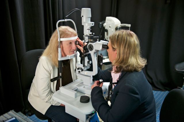
First Bionic Vision Australia patient with her surgeon, Dr Penny Allen.

Dianne Ashworth with her surgeon, Dr Penny Allen. Photo Credit: David Mirabella

Veronika Gouskova, Marketing and Communications Manager of Bionic Vision Australia (BVA) kindly responded by email to my questions about their news:

BSB: I saw in your Aug 30 media release that the implant was described as a “world first” by Dr Allen. Could you clarify in what way this is a “world first” when there are other retinal prosthetics on the market e.g. Argus II from Second Sight?

BVA: This is a world first implantation of a device in the suprachoroidal space at the back of the eye. Although there have been other groups working with patients around the world, this surgical position and procedure is unique to Bionic Vision Australia

BSB: How does the Australian Bionic Eye differ from the Second Sight product that is marketed in Europe?

BVA: There are a number of groups internationally working in the field of retinal implants – the fact that this research is ongoing means that the problem hasn’t yet been solved completely. Between all these groups there are a differences in technology being developed, materials used, surgical placement of the device, surgical technique and the way the electrodes or photodiodes are being stimulated (i.e. how the visual data is processed before it is sent to the implant).

At the end of the day, it’s all about ensuring the best outcome for patients. We are developing two prototypes, with different functional aims: the ‘wide-view’ device combines novel technologies with materials that have been successfully used in other clinical implants. This approach incorporates a microchip with 98 stimulating electrodes and aims to provide increased mobility for patients to help them move safely in their environment.

Our ‘high-acuity’ device incorporates a number of exciting and new technologies, such as diamond materials,  to bring together a microchip and an implant with 1024 electrodes. The device aims to provide functional central vision to assist with tasks such as face recognition and reading large print. The early prototype that we implanted with our first three patients is a stepping stone towards further development for these two devices.

Bionic Vision Australia brings together researchers from many different fields, so we have a truly multidisciplinary team. This means the clinicians and surgeons are involved in the design and development process from the start. Further, a lot of our researchers were involved in the cochlear implant, or bionic ear development – they know what it takes to bring a competitive medical implant to the market.

BSB: I saw that your device was placed between the choroid and sclera, is that significant as opposed to being on the top of the retina? Could you provide further clarification on the significance of this and the surgical technique required for implantation?

BVA: Yes, this early prototype is implanted in the suprachoroidal space, between the choroid and the sclera. This is beneath the retina. There are a number of advantages in doing this, e.g. this position greatly enhances the mechanical stability of the implant and allows for a relatively straightforward surgery. The surgical procedure involves making an incision through the sclera and sliding the implant in place.

BSB: What are the next steps, is a larger clinical trial of your device already planned/underway – when will other patients receive implants?

BVA: We have implanted this early prototype in three patients and will continue to work with these patients over the next 18 months while we further develop our full prototype devices. The next step will be of course, a larger trial with our full devices in due course.

BSB: How might your device be potentially sold/commercialized or made available – is there a commercial partner associated with Bionic Vision Australia?

BVA: Bionic Vision Australia is an unincorporated joint venture between a number of research organisations in Australia. We are funded by the Australian Research Council. A commercialisation vehicle has been set up, Bionic Vision Technologies, to commercialise the technology. This company is solely owned by the member organisations that are involved in our research.

In addition to Second Sight and Bionic Vision Australia, there are several other companies and research groups with retinal prostheses in development.

It is an area where we are seeing innovation in action as teams of multi-disciplinary researchers strive to restore sight and improve the quality of life to people who have become blind.

However, given the high cost of R&D, and the fact that much of the research is government funded, I’m not sure of the commercial opportunity. It will be interesting to see how the market for retinal prostheses develops.

Challenges that have to be overcome

A number of challenges with retinal prostheses will have to be overcome. Some of these are discussed in a 2011 editorial by James Weiland, Alice Cho and Mark Humayun on “Retinal prostheses: Current Clinical Results and Future Needs” that was published in the AAO journal, “Ophthalmology.”  I encourage anyone with an interest in this area to read this insightful review.

Some of the questions I took from this editorial were:

  • Why do some patients respond better than others with an implant?
  • Will retinal remodeling and ongoing degeneration limit the usefulness of implants?
  • What is the best surgical way to ensure optimal placement of the stimulating arrays to maximize visual acuity but avoid problems associated with fixation, and wound closure?
  • Could artificial vision be detrimental to other sensory inputs e.g. the ability of the visual cortex to process data from non-visual Braille reading?

Answers to these and other questions will come as clinical experience is gained and advances in technology improve the design and functionality. In my view this is innovation in action.

Update February 15, 2013 Argus II receives FDA approval

As expected following the positive endorsement of the Ophthalmic Devices Advisory Panel last year, the United States Food and Drug Administration (FDA) announced in a Feb 14, 2013 press release that Second Sight’s Argus II retinal prosthesis system had received approval as a humanitarian use device for the treatment of adults with advanced retinitis pigmentosa.


References

ResearchBlogging.orgHumayun MS, Dorn JD, da Cruz L, Dagnelie G, Sahel JA, Stanga PE, Cideciyan AV, Duncan JL, Eliott D, Filley E, Ho AC, Santos A, Safran AB, Arditi A, Del Priore LV, Greenberg RJ, & Argus II Study Group (2012). Interim results from the international trial of Second Sight’s visual prosthesis. Ophthalmology, 119 (4), 779-88 PMID: 22244176

Weiland JD, Cho AK, & Humayun MS (2011). Retinal prostheses: current clinical results and future needs. Ophthalmology, 118 (11), 2227-37 PMID: 22047893

1 Comment

Biotech Strategy Blog is 1 today!  I can’t believe that a year has gone by so quickly!  Before moving on to year 2, I thought a brief review might be interesting.

What have been the top posts on Biotech Strategy Blog this past year?

In terms of total visitors per post:

  1. Results from NEJM Lucentis v Avastin AMD CATT clinical trial
  2. AUA Results from PIVOT study show no benefit from radical prostatectomy in low risk early stage patients
  3. ASCO 2011 Cabozantinib (XL184) may be an exciting new prostate cancer drug
  4. Merck’s capthepsin-K inhibitor odanacatib in osteoporosis
  5. Update from AACR on new prostate cancer drugs to watch

For those who like metrics:

  • Highest number of reads per month was in May (19,927)
  • Year to date there have been 79,179 visitors
  • Most visited day was September 22, 2011 (2136 reads)

What have been some of the other posts that I enjoyed writing about?

My top 5 (not in rank order) would be:

  1. Alpharadin will be new treatment option for prostate cancer
  2. Patient advocacy session at European Hematology Assocation EHA Congress shows impact of drug adherence on outcome
  3. How nanotechnology may revolutionize the detection of traumatic brain injury using a sensor that changes color
  4. Innovation in Nanotechnology will lead to improved drug delivery, diagnostics & imaging
  5. Insights of the decade

Finally, I have produced 4 videos that you can watch on the biotechstrategy channel on YouTube.

http://youtu.be/nDvY7opm3Fs

http://youtu.be/_oAJ1fU0PT4

http://youtu.be/hM_wmjaqDyc

http://youtu.be/i5GNBmuISqQ
It’s been a busy but enjoyable year. Biotech Strategy Blog is still a work in progress.  If you have enjoyed a particular series of posts or would like me explore a topic or theme in the future, do email me or post a comment.

The FDA earlier this week issued a safety alert to doctors that repackaged bevacizumab (Avastin®) had caused serious eye infections in 12 patients in Florida. The New York Times today reports that five patients at the Veterans Affairs Medical Center in Los Angeles have gone blind as a result of an eye infection following injection of compounded Avastin.

I have written previously about the Lucentis v Avastin debate and the results in the Comparison of Age-related macular degeneration treatment trial (CATT) published earlier this year in the New England Journal of Medicine.

It is not good news that contamination has occurred while compounding bevacizumab (Avastin) from sterile100mg/4mL single use preservative-free vials into individual 1mL syringes.

Genentech/Roche may see this news as reinforcing their position that ranibizumab (Lucentis®) should only be used, since it comes in the correct dose for injection in the eye. However, this ignores the reality caused by the fact that Lucentis is approximately 40x the cost of compounded Avastin ($1950 versus $50).

This week’s news does not support the proposition that intravitreal injection of bevacizumab is not safe and effective for the treatment of AMD, nor any suggestion that pharmacies properly accredited and experienced in aseptic techniques are not qualified to do this. Pharmacists compound drugs everyday.

As the FDA notes in their alert:

“Health care professionals should be aware that repackaging sterile drugs without proper aseptic technique can compromise product sterility, potentially putting the patient at risk for microbial infections.  Health care professionals should ensure that drug products are obtained from appropriate, reliable sources and properly administered.”

However, there is no evidence to suggest that the pharmacies who undertook the Avastin compounding that led to the infection were not “appropriate, reliable sources and properly administered.”

The New York Times notes that the the contaminated Avastin came from the pharmacy at the main campus of the V.A. Greater Los Angeles Healthcare System.  There is no mention of whether the VA pharmacists did the compounding themselves or sourced the drug elsewhere.

According to a news report in the Florida SunSentinel, the pharmacy identified as the source of the infection in Florida is InfuPharma. This is not a retail pharmacist in the high street, but a specialist compounding pharmacy that advertises sterile preparations for numerous products. Licensed pharmacists run this specialist company and looking at their website they do appear to be experienced in this area.

Endophthalmitis is a serious eye infection that may lead to loss of vision. The contamination should not have occurred.  These incidents should not, however, be blown out of proportion in the Lucentis v. Avastin debate.

Sadly, infections and contamination happen in hospitals and the healthcare industry all the time. Even the FDA approved manufacturing facilities of pharmaceutical companies have experienced problems in recent years.  Last summer, BMS experienced issues with sterile manufacturing standards at their Puerto Rico plant following FDA inspections.  Earlier this week, Baxter announced they had filed a lawsuit against Teva for indemnification over a hepatitis C outbreak following reuse of oversize propofol vials.

The news of serious eye infections with repackaged Avastin must, therefore, be put in context. There are countless patients around the world who have benefited from intravitreal injections of Avastin for treatment of their age-related macular degeneration (AMD). The issue raised by the infections in Florida and Los Angeles is whether there is adequate inspection and certification of compounding pharmacies, and whether there is a need for more State regulation and inspections in this area.

Disclosure:  I have written freelance articles for Pharmacy Today, the magazine of the American Pharmacists Association.

1 Comment

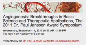
Today, my attention was caught by The New York Academy of Sciences forthcoming seminar on “Angiogenesis: Breakthroughs in Basic Science and Therapeutic Applications.”

Sponsored by Johnson & Johnson and the Dr Paul Janssen Award for Biomedical Research, the seminar (free registration) features some distinguished speakers including Napoleone Ferrara, MD who is giving a presentation on the “Discovery of Anti-Angiogenesis Therapies for Cancer and Ocular Disease.

I’m particularly interested in hearing the panel discussion in the afternoon on the “future of angiogenic medicine,” and the potential of gene transfer therapy as a treatment option.

Those following this area will already know that ocular gene therapy was a topic of discussion at the FDA Cellular, Tissue and Gene Therapies Advisory Committee meeting in June this year.

The briefing document for the meeting discusses how a number of inherited retinal diseases such as Leber Congenital Amaurosis, Stargardt Disease and Retinitis Pigmentosa might benefit from gene therapy.  Given the standard of care is largely supportive in many of these disorders, the potential benefits are huge.

Gene therapy may also offer benefits in the treatment of acquired retinal disorders such as age-related macular degeneration (AMD), the leading cause of blindness in people aged 50 years of older in the developed world.

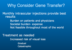
In a presentation to the meeting by Professor Peter Campochiaro of the Wimer Eye Institute at Johns Hopkins, he noted the burden of regular introcular injections.

Potential gene therapy products for retinal disease, such as those using viral vectors and plasmid DNA vectors into which a transgene can be inserted, will be an interesting area to watch.

Due to the pressure of other commitments, I only had the pleasure of attending the annual meeting of the Association for Research in Vision and Ophthalmology (ARVO) for two days, but one of my key take home messages from the meeting is how we can use the eye as a window into the brain.  This is particularly relevant to Alzheimer’s research.

ARVO researchers at a lunchtime workshop that I attended asked the question of what can we learn from shared disease mechanisms in age-related macular degeneration (AMD), Alzheimer’s Disease (AD) and Glaucoma to devise therapies of the future?

What I learnt in the introduction by Nicholas Bazan from LSU Health Sciences is that both AD and AMD are both multifactorial, genetically complex, progressive, late-onset neurodegenerative conditions.  Common features include:

  1. Age-related neurodegeneration
  2. Amyloid precursor protein mis-processing
  3. Non-resolving inflammatory response
  4. Selective apoptotic cell death

Researchers in the workshop presented early experimental findings.

Catherine Bowes Rickman from Duke presented data that showed anti-amyloid immunotherapy blocks retinal pigment epithelium (RPE) damage and visual function defects in an AMD-like mouse model.  Interesting questions were raised as to whether mouse Aß aggregates differently to human, so is this a good model?

Adriana Di Polo from the University of Montreal discussed Glaucoma and AD: common neurodegenerative pathways and therapeutic targets. It was interesting to note that high rates of visual abnormalities, including glaucoma, have been reported in AD patients, but causality has not been established. Neuronal loss in both glaucoma and alzheimer’s disease occurs via common cell death processes including altered metabolism of Amyloid Precursor Protein (APP) and Aß.

What Di Polo highlighted in her talk was the potential to use therapies effective in one disease to treat the other e.g. galantamine is approved for treatment of mild to moderate AD symptoms.  Because it crosses the retinal-brain barrier and has high bioavailability, she presented results using this in an animal model of glaucoma.

Her conclusion was that “therapeutic modalities that promote neuroprotection in AD may be useful in glaucoma and vice versa.”

The third speaker of this fascinating workshop was Ian Trounce from Melbourne, who challenged the Amyloid theory of AD. His hypothesis was that sAPPα may trigger oxidative stress in mitochondria and be the problem. He discussed the increasing acceptance/overlap in pathologies between Parkinson’s and AD.  He presented data that sAPPα overexpression protects retinal ganglion cells (RGC) from rotenone via PI3K-AKT activation.

Critical feedback on the three presentations was provided by Guy Eakin of the American Health Assistance Foundation (AHAF) and Imre Lengyel from UCL.

As Dr Lengyel succinctly notes in his UCL Institute of Ophthalmology bio:

“It appears that the development of age related macular degeneration (AMD) and Alzheimer’s disease (AD) share similar histopathology, vascular risk factors and genetic predisposition. In addition, the development of AMD appears to use similar or identical steps on the cellular and molecular levels to AD: vascular damage, oxidative stress, inflammation, extracellular protein and peptide degradation or deposition, and the role for lipids and trace elements (especially zinc) in the degenerative process are amongst the many common features. Furthermore, amyloid beta peptides are an integral part of drusen (the hallmark lesion in AMD) and their formation might be similar to plaque formation in AD.”

I applaud ARVO for looking at how the eye can be used as a window into the brain. It raises the intriguing prospect that research on AMD may not only help understand the cause of AD, but that the eye may serve as an experimental model for future new treatments. Collaboration between Opthalmology and Alzheimer’s researchers is something I expect and hope we will see more of.


There is a lot of innovative research being presented at the Association for Research in Vision and Ophthalmology (ARVO) annual meeting here in Fort Lauderdale.

For those unable to attend, a large number of poster presentations are now available online with free access.

Not all posters are being made available electronically, only a select few have been invited by the Program Committee, and not every presenter invited has made their poster available.

The online electronic posters will be added to over the next few days of the conference after they have been presented.  I am impressed that ARVO have made this information publicly available at no cost through June 30th.

For those interested the link to the available ARVO 2011 electronic posters can be found on the ARVO website, or you can click here.

The selected posters provide a window into ophthalmology and vision research and are well worth viewing.

4 Comments

This afternoon at the Association for Research in Vision and Ophthalmology (ARVO) annual meeting in Fort Lauderdale saw the long-awaited briefing on the National Eye Institute Lucentis v Avastin comparative effectiveness trial in AMD.

I wrote in a previous post about the CATT results published in the New England Journal of Medicine.

I also tweeted from the briefing, but must add the disclaimer that live tweeting is tough and none of my tweets should be relied upon for accuracy.

What did I learn from the briefing (aside from the fact that ARVO’s technology vendors have real challenges in not being able to light a room properly, set up wifi and get a projector to work):

  • The CATT study was set-up before anyone knew what the price for Lucentis was.
  • No mention was made in the briefing today about comparative cost – but isn’t that what everyone thinks this study is about?
  • No mention was made of how you balance the cost benefits against the trade-off of the higher risk of serious adverse events that was seen.
  • When asked what they would give to a relative, most of the panel answering Q&A “punted” and said they would discuss the data with the patient, and try and get them to make an informed decision.
  • Only Dan Martin from Cleveland Clinic stuck his neck out and said if the patient asked him to make the choice of what to take, he would use Avastin over Lucentis – this statement clearly resonated with the audience (he received a big round of applause)
  • Two year data is already available and some of it was shown today, with no difference in death rate at 2 years between Lucentis or Avastin. The two year data will be published this time next year.
  • The study was not adequately powered to look at the serious adverse events (SAE), which means the statistically significant difference in SAEs may never be truly understood.

Overall, my impression from the briefing is that Lucentis is not identical to Avastin, although functionally the benefits from both appear broadly comparable with no statistical significance between them in terms of visual acuity gains. No mention was made of when you might use one drug over the other (if at all).

Lucentis did have some benefits over Avastin such as a lower retinal thickness at one year compared to the other three treatment groups, which was statistically significant. Both drugs produced an immediate and substantial decrease in retinal fluid, but more eyes were completely dry with Lucentis.

My thoughts are that the debate over Lucentis v Avastin is like the difference between a brand versus a generic, they are similar in terms of efficacy but not identical. While that’s perhaps an over-simplification given that in this case one drug is FDA approved and the other is not, those who want a brand and can afford it will buy a brand, while others may prefer the “generic” for cost reasons.

Will the Lucentis v Avastin decision be any different? If you can afford Lucentis through your insurance you may choose to take that, otherwise Avastin is another option for AMD.  Weighing cost versus slightly higher risk of SAE’s (for which there’s no adequate explanation showing causality) is how I would approach this decision.

It was interesting to note that the follow-up will continue and two year results will be published this time next year. They may shed further light on the serious adverse event differences, but I suspect that given the way the study is powered, this issue may not be resolved any further.

My overall take is that there is nothing compelling in the data to suggest that any ophthalmologist that is using bevacizumab off-label will switch patients to ranibizumab. Off-label Avastin use in wet AMD received an official NEI endorsement from the CATT study and will continue.

Equally those who are happy with Lucentis will not switch to Avastin on the back of this study, given that the Lucentis data appeared slightly better in places.  The controversy will no doubt continue and other studies comparing Lucentis v Avastin will add further insight.


The annual meeting of the Association for Research in Vision and Ophthalmology (ARVO) starts tomorrow in Fort Lauderdale. There are some educational courses that are also running today.
Many of the vision researchers and ophthalmologists will be sharing their thoughts from the conference.

If you are interested in following the Twitter discussion (hashtag #ARVO11), you can do so using the aggregator below – just click on the play button to see the tweets.

2 Comments

There is a lot of buzz this week about Lucentis versus Avastin for the treatment of wet age-related macular degeneration (AMD), something that will be talked about in more detail at the Association for Research in Vision and Ophthalmology (ARVO) annual meeting this weekend in Fort Lauderdale.

Also on the radar at ARVO is more news from Second Sight and their Argus II Retinal Prosthesis (something that I have previously written about on this blog).  For those interested there is a press conference at ARVO on Tuesday, May 3 from 5-6pm.

Second Sight presents updated results from the Argus II Retinal Prosthesis clinical trial, including sentence reading and color vision restoration for previously blind subjects. Two trial participants and independent investigators from the trial will be available for interviews.

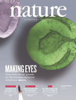
Which brings me back to a Nature article published earlier this month that I have been meaning to write about showing, for the first time, the ability to generate a three-dimensional culture of neural retinal tissue from mouse embryonic stem (ES) cells.  A word of warning, you may find the paper a little tough to follow unless you are a scientist in this field.

Eiraku and colleagues from Japan were able to culture retinal tissue similar to that seen in the human eye.  Eye formation starts as an optical vesicle that then develops into a two-walled optic cup.  As the authors note “optic cup development occurs in a complex environment affected by neighbouring tissues.”

What the authors showed in their research was the ability to culture retinal tissue containing ganglion cells, photoreceptors and bipolar cells.  They conclude:

Collectively, these findings demonstrate that the fully stratified neutral retina tissue architecture in this ES-cell culture self-forms in a spatiotemporally regulated manner mimicking in-vivo development.

My take on this research is that it is an important milestone in regenerative medicine that could lead to the prospect of retinal transplants in the future.  I look forward to learning more at ARVO about what the future may hold for retinal transplants derived from human stem cells.

ResearchBlogging.orgEiraku, M., Takata, N., Ishibashi, H., Kawada, M., Sakakura, E., Okuda, S., Sekiguchi, K., Adachi, T., & Sasai, Y. (2011). Self-organizing optic-cup morphogenesis in three-dimensional culture Nature, 472 (7341), 51-56 DOI: 10.1038/nature09941

Breaking news:

The New England Journal of Medicine have just published online the results of the Comparison of Age-related macular degeneration treatment trial (CATT) comparing the efficacy of FDA approved ranibizumab (Lucentis) to off-label bevacizumab (Avastin); a trial that has important commercial importance given the comparative costs of an intravitreal injection of around $1950 (Lucentis) vs. $50 (Avastin).

Key Study Results

Based on 1208 patients randomly assigned in the single-blind noninferiority trial, primary outcome was mean change in visual acuity between baseline and 1 year. This was equivalent between the two drugs.

Bevacizumab administered monthly was equivalent to ranibizumab administered monthly, with 8.0 and 8.5 letters gained, respectively.

Secondary outcome measures included the incidence of ocular and systemic side effects, the results show some similarities and differences:

Rates of death, myocardial infarction and stroke were similar for patients receiving either bevacizumab or ranibizumab (P>0.20).

The proportion of patients with serious adverse events (primarily hospitalization) was higher with bevacizumab than with ranibizumab (24.1% vs 19%)

The conclusion of the study is that:

At 1 year, bevacizumab and ranibizumab had equivalent effects on visual acuity when administered to the same schedule”

However, here is the potential ‘get out’ for Genentech:

Differences in rates of serious adverse events require further study.

The investigators note that the difference in serious adverse events may be due to:

“chance, imbalances in baseline health status that were not included in the medical history or multivariate models, or a true difference in risk.”

i.e. they don’t know.

What the results from the CATT study mean is that Avastin and Lucentis are similar, but different. That is not a surprising result given that they originate from the same anti-VEGF monoclonal antibody.  However, they are not identical.

Clearly, if I were a patient, the additional 5% risk of serious adverse events would have to be weighed against the cost benefits. For those who are uninsured or unable to afford Lucentis, receiving Avastin may be an informed decision worth taking.  As the investigators note:

One of the many factors that contribute to the selection of a drug for a patient is cost.  A single dose of ranibizumab costs 40 times as much as a single dose of bevacizumab.  This cost differential has important economic implications when extrapolated to the more than 250,000 patients who are treated for neovascular AMD annually in the United States.

I look forward to hearing the animated discussion of these results at the ARVO annual meeting in Fort Lauderdale on Sunday.

ResearchBlogging.orgThe CATT Research Group (2011). Ranibizumab and Bevacizumab for Neovascular Age-Related Macular Degeneration New England Journal of Medicine DOI: 10.1056/NEJMoa1102673

1 Comment
error: Content is protected !!