Biotech Strategy Blog

Commentary on Science, Innovation & New Products with a focus on Oncology, Hematology & Cancer Immunotherapy

Posts from the ‘Tissue Engineering’ category

Credit: Silvia A Ferreira, Cristina Lopo and Eileen Gentleman, KCL

Recently a reader wrote in asking if there were any particular cell therapy products in early development that might be worth watching out for or exploring ahead of time that I would recommend following?

The approach described here fits that category nicely and also has the added bonus of potentially having multiple applications and use cases.

Some of them are in the non-oncology space too.

There were some interesting insights that emerged during this research that I thought were well worth sharing…

To learn more from our latest analysis and get a heads up on our oncology insights, subscribers can log-in or you can click to gain access to BSB Premium Content.

This content is restricted to subscribers

Regular blog readers will know I think tissue engineering is an exciting area where you can see innovation in action – advances in basic science can translate into ways to artificially create replacement organs and body parts.

Research published online 22 July 2012 in Nature Biotechnology by Janna Nawroth and colleagues at the California Institute of Technology (Caltech) and Harvard University, shows how biomedical engineers are learning from the structure and function of other animals.

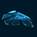
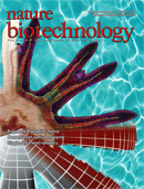
In a Nature Biotechnology article titled “a tissue-engineered jellyfish with biomimetic propulsion” researchers describe how they were able to combine rat cardiac muscle cells and a synthetic elastomer membrane into a medusoid like structure that mimicked the propulsion of a jellyfish.

Credit: Caltech and Harvard University

They achieved this by forming the elastomer into a medusoid or jellyfish like shape with eight lobes around a central disc, then applying a monolayer of rat cardiac muscle tissue, which when electrically shocked, contracted in a synchronized way.

The net result was the medusoid “swam” in a similar way to a jellyfish. They effectively developed an artificial pump made out of a hybrid of living cells and silicone rubber.

The video below by Janna Nawroth, produced by Caltech and Harvard University, shows the medusoid in action, and explains how this research advances the design of muscular pumps for biomedical application:

According to the Caltech press release, this approach in reverse-engineering the function of a jellyfish “will be broadly applicable to the reverse engineering of muscular organs in humans.” 

While we are not yet able to tissue engineer a replacement human heart, it’s hard not to believe that at some point in the future we will see the development of hybrid devices that combine synthetic materials and cultured heart muscle cells.

Reference

ResearchBlogging.orgJanna C Nawroth, Hyungsuk Lee, Adam W Feinberg, Crystal M Ripplinger, Megan L McCain, Anna Grosberg, John O Dabiri, & Kevin Kit Parker (2012). A tissue-engineered jellyfish with biomimetic propulsion Nature Biotechnology, 30, 792-797 DOI: 10.1038/nbt.2269

Biotech Strategy Blog is 1 today!  I can’t believe that a year has gone by so quickly!  Before moving on to year 2, I thought a brief review might be interesting.

What have been the top posts on Biotech Strategy Blog this past year?

In terms of total visitors per post:

  1. Results from NEJM Lucentis v Avastin AMD CATT clinical trial
  2. AUA Results from PIVOT study show no benefit from radical prostatectomy in low risk early stage patients
  3. ASCO 2011 Cabozantinib (XL184) may be an exciting new prostate cancer drug
  4. Merck’s capthepsin-K inhibitor odanacatib in osteoporosis
  5. Update from AACR on new prostate cancer drugs to watch

For those who like metrics:

  • Highest number of reads per month was in May (19,927)
  • Year to date there have been 79,179 visitors
  • Most visited day was September 22, 2011 (2136 reads)

What have been some of the other posts that I enjoyed writing about?

My top 5 (not in rank order) would be:

  1. Alpharadin will be new treatment option for prostate cancer
  2. Patient advocacy session at European Hematology Assocation EHA Congress shows impact of drug adherence on outcome
  3. How nanotechnology may revolutionize the detection of traumatic brain injury using a sensor that changes color
  4. Innovation in Nanotechnology will lead to improved drug delivery, diagnostics & imaging
  5. Insights of the decade

Finally, I have produced 4 videos that you can watch on the biotechstrategy channel on YouTube.

http://youtu.be/nDvY7opm3Fs

http://youtu.be/_oAJ1fU0PT4

http://youtu.be/hM_wmjaqDyc

http://youtu.be/i5GNBmuISqQ
It’s been a busy but enjoyable year. Biotech Strategy Blog is still a work in progress.  If you have enjoyed a particular series of posts or would like me explore a topic or theme in the future, do email me or post a comment.

BioPharm America 2011 Banner

A conference I regretably will not be at, but would have like to have attended is BioPharm America 2011 – 4th International Biotechnology Partnering Conference that is taking place in Boston from today until this Friday, September 9th.

The program overview suggests that it will be an interesting meeting with sessions on personalized medicine, business development and strategy and partnering. On friday there’s a briefing on Regenerative Medicine and Cell Therapy: The Road to Commercialization. If like me, you are unable to attend, you can follow the conversation on twitter using the hashtag #BPA11 (nice and short!).  I noticed there’s already some excellent live tweeting from the event. I’ve added an aggregator below to make it easier to follow or catch up on the news. Just click on the play button to see the tweets:  

I grew up watching Lee Majors in the 1970’s TV show “The Six Million Dollar Man”, about an injured former astronaut whose bionic implants allowed him to do superhuman feats.

That was fiction, but it is becoming closer to reality as a result of new research into how artificial limbs can integrate with human tissues. This work on neural-electrical interfaces may ultimately allow someone to control a prosthesis as you would a normal limb, i.e. by nerve impulses that travel from the brain.

The promise of “functional integration” between the human nervous system and an electro-mechanical device is the greater control this would give amputees and potential for sensory feedback.

The laboratory of D. Kacy Cullen, Ph.D, assistant professor in the Department of Neurosurgery, Center for Brain Injury & Repair at the University of Pennsylvania, Perelman School of Medicine is actively working in this area. I had the pleasure to hear Dr Cullen talk about his research at Health Journalism 2011 earlier this year. I previously wrote on this blog about his research on nanomaterials that change color with blast impact.
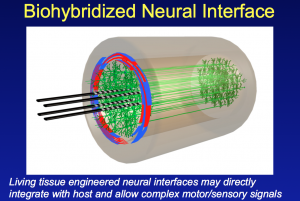
Source: D Kacy Cullen PhD, University of Pennsylvania

In his presentation to the Association of Health Care Journalists he described some of the neural tissue engineering work in his laboratory. This research has shown the ability to integrate axons in the peripheral nervous system (PNS) with an array of electrodes embedded in a living collagen matrix.

In essence this is the development of a nerve tissue/electrical interface that potentially allows the integration of man and machine.
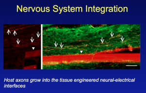
Nervous System Integration


Key to success has been the development of a living, 3-D scaffold where this integration between nerve and electrodes can take place. Nerve axons in the peripheral nervous system require a living target for innervation. This has involved designing and engineering a 3-D living cellular matrix/scaffold that will work within a living body (to date the research has been on animal models).

Not only is Cullen and his laboratory looking at machine/nerve interfaces, but they have also developed techniques that may allow nerves to be repaired.  They have shown that nerves can be elongated or stretched using novel tissue engineering techniques.

The resulting axonal constructs via stretch-growth have been transplanted into rats and used to bridge an excised segment of sciatic nerve.  What was subsequently seen was an interwining plexus of host and graft axons, suggesting axonal regeneration across the lesion.

While still early stage, and not yet tested in humans, this research has tremendous potential for those paralysed due to traumatic nerve damage in the future.

Moving forward, the Penn researchers aim to develop an application for CNS tissue repair that can be delivered to the brain or spinal cord via stereotactic microinjection. This is minimally invasive surgery that allows delivery of cells to a precise location using 3D co-ordinates.

According to research published in Critical Reviews™ in Biomedical Engineering they plan to use micro-engineered hydrogel conduits, several centimeters long and the width of three hairs. These hydrogels will contain living axonal tracts that will then hopefully reconnect damaged nerves, and provide a platform or path for regeneration.

Many challenges still remain to be worked out for the tissue engineered neural constructs such as issues relating to inflammation and immune tolerance.

The research also needs to move from animals to humans, and be shown to be safe. The long-term functional outcome is also unknown.  It is far too early to think of this as a treatment option.

However, advances in neural tissue engineering, neuroregeneration and neuro-prosthetics do offer a lot of promise and hope to the many patients who suffer from spinal cord injuries or loss of a limb.

In a short blog post, I have not been able to do full justice to the innovative research or fully describe the techniques and methodology.  More information on the fascinating work being done at Penn, along with details of the associated scientific publications, can be found on the web page of the Cullen Laboratory: Neural Engineering in Neurotrauma.

ResearchBlogging.orgD. Kacy Cullen, John A. Wolf, Douglas H. Smith, & Bryan J. Pfister (2011). Neural Tissue Engineering for Neuroregeneration and Biohybridized Interface Microsystems In vivo (Part 2) Crit Rev Biomed Eng., 39 (3), 243-262
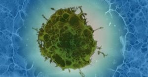

Regenerative Medicine and the science behind replacing body parts with synthetic tissue engineered versions took another step forwards today after researchers announced they had transplanted a trachea made of a nanomaterial covered with the patient’s own cells.

Professor Seifalian and Clare Crowley UCL

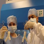
Researchers from University College London led by Prof. Alexander Seifalian designed and built a polymer based nanocomposite tracheal scaffold, which was then seeded with the patient’s own stem cells.

After two days in a bioreactor (Harvard Bioscience), the cells and the synthetic trachea scaffold were transplanted last month at the Karolinska University Hospital in Stockholm by Prof. Paolo Macchiarini and colleagues, into a patient with late stage tracheal cancer.

As reported by BBC health, and the press releases of University College London (UCL), Karolinska Institute and Harvard Bioscience, the 36 year old man is doing well and because the cells on the trachea were his own, no immunosuppressive drugs were needed.

In the UCL press release, Professor Seifalian said:

“What makes this procedure different is it’s the first time that a wholly tissue engineered synthetic windpipe has been made and successfully transplanted, making it an important milestone for regenerative medicine. We expect there to be many more exciting applications for the novel polymers we have developed.”

While this is still experimental research that needs to be validated in a clinical trial with more subjects, there is the potential for Professor Seifalian’s nanomaterial based tissue scaffold to be used for commercial medical devices such as coronary stents and grafts.

In addition to the development of a nanomaterial that can be used as a tissue scaffold, key to success of the transplant was the ability to grow and cover the engineered material with the patient’s own stem cells.  Harvard Bioscience have specifically designed a bioreactor to culture cells onto a graft for airway tissue reengineering.

As innovation in science drives new milestones in regenerative medicine, we can expect the market for tissue engineered products to grow as companies seek regulatory approval for commercial products.

Above all else, regenerative medicine offers major benefits for patients and the restoration of function and improved quality of life. Today’s news is yet another milestone that highlights the promise of regenerative medicine.

BIO-2011-Interational-Convention-Washington-DC

I am excited to be attending, for the first time, the Biotechnology Industry Organization (BIO) international convention that takes place in Washington DC in just over a week’s time from Monday June 27 to Thursday, June 30th.

This meeting has something for everyone interested in the biotechnology industry whether it be deal making, partnering, licensing, drug discovery or personalized medicine. There are 16 specialized tracks where industry experts provide insight and best practices.

In addition, there are numerous networking and social events plus an exhibit hall that showcases the world’s biotech regions and how they are promoting innovation.

At meetings where there are parallel sessions, I apply “the law of two feet” (thanks to Podcamp for this) that says if you are not getting what you want from the session, it’s OK to walk out and go to another one.

My top 10 sessions at BIO reflect my personal interests in innovation, science and new product development:

Tuesday June 28

  • How will we afford Personalized Medicines?
  • The Biomarkers Consortium: Facilitating the Development and Qualification of Biological Markers
  • Personalized Oncology: The emergence of Personalized Medicine Strategies in Oncology Clinical Development and Deal Making
  • Navigating the New Law on Licensing Biosimilars

Wednesday June 29

  • Lessons from a Mature Public-Private Partnership. The Alzheimer’s Disease Neuroimaging Initiative
  • Emerging Markets. The Future of Growth for Biologics?
  • The Role of Imaging Biomarkers in Early Phase CNS Drug Development
  • The Promise of MicroRNA-based Therapeutics in Cancer

Thursday Jun 30

  • After the Fall. Venture Capital and the Biotech Funding Landscape
  • Regulatory Issues for Tissue Engineered Products

If you have plans to be at BIO 2011 do say hello after one of the sessions or receptions. You can reach me at the meeting via twitter (@3NT).  See you in DC!

Follow 3NT on Twitter

1 Comment

There is a lot of buzz this week about Lucentis versus Avastin for the treatment of wet age-related macular degeneration (AMD), something that will be talked about in more detail at the Association for Research in Vision and Ophthalmology (ARVO) annual meeting this weekend in Fort Lauderdale.

Also on the radar at ARVO is more news from Second Sight and their Argus II Retinal Prosthesis (something that I have previously written about on this blog).  For those interested there is a press conference at ARVO on Tuesday, May 3 from 5-6pm.

Second Sight presents updated results from the Argus II Retinal Prosthesis clinical trial, including sentence reading and color vision restoration for previously blind subjects. Two trial participants and independent investigators from the trial will be available for interviews.

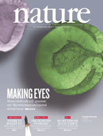
Which brings me back to a Nature article published earlier this month that I have been meaning to write about showing, for the first time, the ability to generate a three-dimensional culture of neural retinal tissue from mouse embryonic stem (ES) cells.  A word of warning, you may find the paper a little tough to follow unless you are a scientist in this field.

Eiraku and colleagues from Japan were able to culture retinal tissue similar to that seen in the human eye.  Eye formation starts as an optical vesicle that then develops into a two-walled optic cup.  As the authors note “optic cup development occurs in a complex environment affected by neighbouring tissues.”

What the authors showed in their research was the ability to culture retinal tissue containing ganglion cells, photoreceptors and bipolar cells.  They conclude:

Collectively, these findings demonstrate that the fully stratified neutral retina tissue architecture in this ES-cell culture self-forms in a spatiotemporally regulated manner mimicking in-vivo development.

My take on this research is that it is an important milestone in regenerative medicine that could lead to the prospect of retinal transplants in the future.  I look forward to learning more at ARVO about what the future may hold for retinal transplants derived from human stem cells.

ResearchBlogging.orgEiraku, M., Takata, N., Ishibashi, H., Kawada, M., Sakakura, E., Okuda, S., Sekiguchi, K., Adachi, T., & Sasai, Y. (2011). Self-organizing optic-cup morphogenesis in three-dimensional culture Nature, 472 (7341), 51-56 DOI: 10.1038/nature09941

There are 5,396 posters at the 102nd Annual Meeting of the American Association for Cancer Research (AACR) here in Orlando. Intermingled with the exhibitors (something that no doubt encourages traffic to the exhibits), the posters provide a window into the world of current cancer research and the spirit of collaboration.
Researchers from all over the world present their latest scientific discoveries, what they may have spent 3 years or more years on while studying for a Ph.D or undertaking a post-doctoral fellowship.

The research is innovative, and what’s seen at AACR is often at the cutting edge and shown prior to publication in a major journal.

What is palpable is the energy surrounding the poster discussions as experts, thought leaders and leading researchers network and share ideas with typically more junior colleagues, and in the process relate their experience to the poster being presented.

In a world of fixed term grants, the poster session is also an opportunity to showcase research to those who may be looking to hire new talent to their team.

It takes six poster sessions over four days for the 5000+ posters to be presented. I’m looking forward to the exercise!

Taxanes are a class of drug that are used in breast, lung and ovarian cancer chemotherapy to disrupt the function of microtubules that are essential to cell division. They include paclitaxel (Taxol®) and docetaxel (Taxotere®).

Paclitaxel is also used to prevent the narrowing (restenosis) that occurs with coronary artery stents that are used to open blocked coronary arteries. Drug coated stents (a.k.a. “drug-eluting stents) reduce scar tissue.

Research published in the February 18, 2011 edition of Science, by Farida Hellal and colleagues has now shown that treatment with paclitaxel reduces the scarring associated with spinal cord injury (SCI) and promotes nerve regeneration.

The paper in Science is well worth reading and takes the reader through a logical thought process as the researchers tested their hypothesis that paclitaxel might stabilize microtubules around the site of SCI.

One of the cellular events that occurs after SCI is the activation of transforming growth factor-ß signaling (TGF-ß).

Increased TGF-ß leads to fibrosis or scarring.  TGF-ß acts on Smad2 to bind to microtubules through kinesin-1.  Hellal and colleagues asked if treatment with paclitaxel would impair Smad-dependent TGF-ß signaling? The answer from their elegant series of experiments is that yes it does.

Not only that, but TGF-ß also regulates the axon growth inhibitor, chondroitin sulfate proteoglycans (CSPGs).  The researchers asked whether pacllitaxel decreased CSPGs after SCI?  They found that cultured meningeal cells and astrocytes treated with 10 nM paclitaxel showed a 35% and 32% decrease of glycosaminoglycan (GAG) levels.

The next logical question is whether the reduction of scar formation by paclitaxel results in any benefits for new nerve formation? The regeneration of dorsal root ganglions (DRG) were evaluated.  In what to me was a finding of great significance, the researchers found (references to figures omitted) that:

“Taxol-treated animals had regenerative fibers growing along the edge of the lesion cavity into the injury site and beyond. The longest axons per animal grew 1199 T 250 mm in the Taxol-treated group versus 176 T 225 mm in the vehicle-treated an- imals (n = 13 animals per group; P = 0.002; two- tailed t-test). The Taxol-treated lesion site thus becomes favorable for regeneration of growth-competent axons.”

The final part of this research asked whether treatment with paclitaxel led to any functional improvement after the test animals received a spinal cord injury? They found that those rats that received paclitaxel after injury, had greater improvement in their locomotor function.   The conclusion being that “Taxol-induced functional recovery correlates with its axon growth–inducing effect.”

The results from any animal study must be viewed with caution, since they don’t necessarily translate to humans.  However, this animal research, if supported by data from human clinical trials, suggests that treatment with taxanes may be of benefit to those with spinal cord injuries.

Given the debilitating effect of any spinal cord injury, this is an important finding.


ResearchBlogging.orgHellal, F., Hurtado, A., Ruschel, J., Flynn, K., Laskowski, C., Umlauf, M., Kapitein, L., Strikis, D., Lemmon, V., Bixby, J., Hoogenraad, C., & Bradke, F. (2011). Microtubule Stabilization Reduces Scarring and Causes Axon Regeneration After Spinal Cord Injury Science, 331 (6019), 928-931 DOI: 10.1126/science.1201148

error: Content is protected !!